Periotest
dynamically diagnosing the human periodontium and the dental
implant-bone
interface
 about
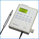
Periotest device
about
Periotest device
This web site is an uncommercial one.
Though the contents have been accurately verified, no warranty may
be issued.
Only the authors are responsible for the content of their papers.
References: ...
1989
1990 1991
1992 1993
1994 1995
1996 1997
1998 1999
2000 2001
2002
2003
2004 2005 2006 ...........
2006:
Resonance frequency analysis and
damping capacity assessment. Part I: an in vitro study on measurement
reliability and a method of comparison in the determination of primary
dental implant stability
LACHMANN S, JAGER B, AXMANN D, GOMEZ-ROMAN G, GROTEN M, WEBER
H., Department of Prosthodontics, University of
Tubingen, Tubingen, Germany. stefan.lachmann@med.uni-tuebingen.de
Clin Oral Implants Res. 2006 Feb;17(1):75-9.
Evaluation Study
OBJECTIVES: The aims of this in vitro study were to evaluate reliability of the Osstell and Periotest
devices in the assessment of implant stability and to perform a method
comparison. MATERIAL AND METHODS: Commercial dental implants were
inserted into bovine rib segments of different anatomical origins and
densities. Repeated measurements were performed, varying (a) the
torque-in force of the devices' attachment screw (the Osstell
transducer and the ball attachment, insert for the Periotest
device), (b) the insertion site bone quality, and (c) the thread
exposure in simulated peri-implant bone defects. RESULTS: Both methods
were comparably reliable and showed a strong association to each other
in the classification of implant stability. As opposed to torque-forced
screw attachment, the variations in bone composition, differences in
inter-implant stability of adjacent implants, and peri-implant bone
reduction were statistically significant for both methods. CONCLUSIONS:
Both non-invasive diagnostic devices seem to be useful in the long-term
follow-up of implant integration.
Resonance
frequency analysis and damping capacity assessment. Part 2:
peri-implant bone loss follow-up. An in vitro study with the Periotest
and Osstell instruments
LACHMANN S, LAVAL JY, JAGER B, AXMANN D, GOMEZ-ROMAN G, GROTEN M, WEBER
H., Department of Prosthodontics, University of
Tubingen, Tubingen, Germany. stefan.lachmann@med.uni-tuebingen.de
Evaluation Study, Historical Article
OBJECTIVE: We compared the performance of damping capacity assessment (Periotest
device) to resonance frequency analysis (Osstell device) in the
assessment of peri-implant bone loss in an in vitro experiment.
MATERIAL AND METHODS: Screw-type oral implants were polymerized into
acrylic blocks. Peri-implant bone loss was simulated by successively
removing defined portions of material surrounding the implants in
millimeter increments. Measurement values of both devices were compared
by assessing the associated measurement errors, by calculating
correlation analyses and drawing scatterplots, and by means of
regression analysis referring to increasing bone loss. RESULTS: Both
devices produced comparable results suggesting agreement of the
measured implant stability values to the actual loss of peri-implant
resin. There was a noticeable correlation of the Periotest
and Osstell implant stability values. CONCLUSION: The results of this
experiment suggest agreement in predicting the actual implant stability
with both the instruments with the Osstell instrument being the more
precise device.
Implant-borne prosthetic rehabilitation of bone-grafted cleft versus traumatic anterior maxillary defects
LANDES CA., Maxillofacial and Plastic Facial Surgery Department,
The J.-W. Goethe University Medical Centre, Frankfurt, Germany.
c.landes@lycos.com
J Oral Maxillofac Surg. 2006 Feb;64(2):297-307.
PURPOSE: This study hypothesizes comparable implant success in
bone-grafted cleft-alveolus versus traumatic anterior maxillary
defects. Though of different pathogenesis, both defects comprise bone
deficit, scarred periosteum, and soft tissues. Additional complicating
factors are isolated. PATIENTS AND METHODS: Twenty cleft and 20
traumatic defect cases were followed-up 48 months in average. After 9
secondary and 11 tertiary cleft-osteoplasties, 25 implants were
inserted; in traumatic defects following 8 two-stage and 12 one-stage
osteoplasties, 37 incisor or canine implants were inserted. After
secondary and tertiary cleft-osteoplasties, 57 and 13 months elapsed
until implantation, 4 months in the two-stage posttraumatic
osteoplasties. Implants were loaded at 6 months by single crowns.
RESULTS: Four (20%) cleft patients faced 2 failures and 2 first-year
losses; 2 (10%) trauma cases faced 2 failures and 2 first-year losses;
and cumulative 5-year implant success was 80% and 88%, respectively.
Other parameters' 12-month results were: values for mean cleft, trauma
patients (+ standard deviation), significance of comparison in t testing at a cut-off level of alpha = 0.05; bone loss 0.3+0.5 mm, 0.5+0.7 mm, P < .2; Periotest score 1.1+3.1, 1.2+2.5, P < .7; gingival recession 2.1+0.3 mm, 2.2+0.5 mm, P < .6; periimplant probing depth 2.5+0.5 mm, 2.8+2.6
mm, P < .3. CONCLUSION: Similar success rates without statistically
significant differences were found; a multiple factor analysis
discerned as positive predictive factors the following; generous
transplant volume, 3 to 6 months latency, sufficient implant dimension,
early adulthood. Early loading cannot be encouraged from the success
rates. Negative predictive factors were spongiosa or milled-bone
transplants, dehiscence, smoking, and anorexia. Intraorally harvested
membranous bone transplants may prospectively amend secondary
osteoplasty-associated bone resorption. Donor site morbidity, local
growth, and tooth breakthrough require additional observation in a
prospective study when implant insertion should directly follow the
growth spurt.
Outcomes of fixed prostheses supported by immediately loaded endosseous implants.
TORTAMANO P, ORII TC, YAMANOCHI J, NAKAMAE AE, GUARNIERI TDE C.,
Faculdade de Odontologia da Universidade de Sao Paulo, Brazil.
tortamano@giro.com.br
Int J Oral Maxillofac Implants. 2006 Jan-Feb;21(1):63-70.
Evaluation Study
PURPOSE: The aim of this article was to evaluate the survival and
success of Straumann implants after immediate loading. A new method for
fabricating effective definitive prostheses to immediately load
implants in edentulous patients was presented. MATERIALS AND METHODS:
Nine patients received 4 implants each, and resin-metal prostheses were
installed less than 48 hours after implant placement. Mobility was
evaluated immediately after the surgical procedures and 3 months
subsequently using the Periotest.
Clinical evaluation of soft peri-implant tissues was conducted monthly
after the sutures were removed, and radiographs were obtained 6, 12,
and 24 months after the surgery. RESULTS: The Periotest
revealed statistical values that were stable, with no mobility. No
signs of inflammation and/or bleeding were observed. The radiographs
did not reveal any continuous areas of radiolucency beyond the first
thread of the 36 implants after 24 months. None of them failed, and the
success rate was 100%. DISCUSSION: It is possible to submit implants to
immediate load without jeopardizing osseointegration if parameters are
met, such as suitable bone quality and quantity, lack of unfavorable
systemic and psychologic factors, lack of parafunctional habits, strict
maintenance of prosthetic requirements, minimization of micromotion,
and use of an appropriate surgical protocol. CONCLUSION: Under
immediate load, osseointegration of implants is possible, and the
method presented for the fabrication of resin-metal prostheses has been
reliable and predictable.
Implant-supported mandibular overdentures retained with ball or telescopic crown attachments: a 3-year prospective study
KRENNMAIR G, WEINLANDER M, KRAINHOFNER M, PIEHSLINGER E., Department of
Prosthodontics, Dental School, University of Vienna, Austria.
krennmair@aon.at
Int J Prosthodont. 2006 Mar-Apr;19(2):164-70.
Randomized Controlled Trial
PURPOSE: The aim of the present study was to evaluate implant
survival, peri-implant conditions, and prosthodontic maintenance
requirements for implant-supported mandibular overdentures in atrophic
mandibles retained with ball or resilient telescopic crown attachments
during a 3-year period. MATERIALS AND METHODS: Twenty-five patients
with edentulous mandibles each received 2 Camlog root-form dental
implants in the mandibular interforaminal (canine) region. The denture
attachment system was chosen randomly; 13 patients received ball
attachments and 12 patients received resilient telescopic crowns.
Implant survival, implant mobility (Periotest
values), and peri-implant conditions such as bone resorption, pocket
depth, Plaque Index, Gingiva Index, Bleeding Index, and Calculus Index
values were assessed for each implant. In addition, detailed
prosthodontic maintenance was evaluated during the follow-up period and
the 2 retention modalities were compared. RESULTS: There were no
differences in implant survival, implant mobility (Periotest
values), and peri-implant conditions between the 2 retention
modalities. During the 3-year period significantly more
complications/interventions for maintenance purposes were registered in
the ball group (62 interventions) than in the telescopic crown group
(26 interventions; P < .01). CONCLUSION: The results indicate that
both ball attachments and resilient telescopic crowns used on isolated
implants in the edentulous mandible are viable treatment options.
Implant success and peri-implant conditions did not differ between ball
attachments and telescopic crowns used as retention modalities for
implant overdentures, but the frequency of technical complications was
significantly higher with ball attachments than with resilient
telescopic crowns.
Occlusal interference during mastication can cause pathological tooth mobility.
ISHIGAKI S, KUROZUMI T, MORISHIGE E, YATANI H., Department of Fixed
Prosthodontics, Osaka University Graduate School of Dentistry, Osaka,
Japan. ishigaki@dent.osaka-u.ac.jp
J Periodontal Res. 2006 Jun;41(3):189-92.
BACKGROUND AND OBJECTIVE: Despite little evidence regarding the
relationship between tooth mobility and nonworking contact, the
evaluation of occlusion is performed mainly by the detection of
premature and/or nonworking contacts during tapping movements and
lateral excursion. The hypothesis of this study is that occlusal
contact during mastication is potentially traumatic to periodontal
tissue. It clarifies the relationship between chewing patterns and the
status of periodontal tissue. MATERIAL AND METHODS: Subjects included
73 adults, 20-29 years of age (39 men and 34 women), with complete sets
of teeth and no history of orthodontic treatment or periodontal
disease. The closing chewing patterns of each subject were classified
into three groups by the Masticatory Deviation Index, which depicts the
deviation from the normal chewing patterns within 5 mm from the
intercuspal position. Periotest
was used to diagnose teeth mobility and the values were compared among
the three groups. RESULTS: The present study indicates that the chewing
movements which deviated from the normal chewing movements increased
the mobility of specific types of teeth. CONCLUSION: The results of
this study imply a relationship between chewing movements and tooth
mobility and indicate that functional evaluation of occlusion is
necessary for the examination of periodontal tissue. Occlusal
evaluation with border and tapping movements might be insufficient, and
functional occlusal evaluation during chewing movements can be
clinically useful for using to evaluate periodontal tissue.
Comparison of early telescope loading of non-submerged ITI implants in irradiated and non-irradiated oral cancer patients.
LANDES CA, KOVACS AF., Maxillofacial and Plastic Facial Surgery, The
J.-W. Goethe, University Medical Centre, Frankfurt, Germany.
c.landes@lycos.com
Clin Oral Implants Res. 2006 Aug;17(4):367-74.
OBJECTIVE: To compare early dental implant loading in irradiated and
non-irradiated oral cancer patients in order to accelerate masticatory
function improvement and quality of life. PATIENTS AND METHODS: One
hundred and fourteen non-submerged interforaminal ITI implants were
early loaded in 30 patients after 3 weeks in situ (telescoped
overdenture). Nineteen patients received 72 implants (63%) after local
irradiation; 11 non-irradiated patients received 42 implants (37%) with
a 24-month follow-up. RESULTS: At 24 month follow-up, one early failure
had occurred in an irradiated patient (=99% functioning implants in
situ). Peri-implant bleeding and plaque index were similarly high in
both groups (40 to 68% average).The Results of other measured
parameters were as follows (values for mean; irradiated; non-irradiated
patients with respective standard deviations; significance of
comparison): bone loss (0.9+/-0.9; 1.4+/-0.9; 0.4+/-0.5 mM;
P<0.01); Periotest
score (-2.7+/-2.7; -2.4+/-2.2; -3.1+/-3.3; P<0.2); gingival
recession (0.6+/-0.7 mM; 0.8+/-0.9 mM; 0.4+/-0.5 mM, P<0.02); and
peri-implant probing depths (3+/-1.2; 2.6+/-0.6; 3.4+/-1.7 mM;
P<0.002). CONCLUSION: The results suggest reliable non-submerged
implantation and early loading. However, bone loss in irradiated
mandibles, combined with higher average Periotest
values and gingival recession in an oral environment of altered saliva
quantity, quality, microflora and local scarring, requires extended
follow-up.
Early implant failure. Prognostic capacity of Periotest: retrospective study of a large sample
NOGUEROL B, MUNOZ R, MESA F, DIOS LUNA J, de, O'VALLE F., Department of
Periodontology, School of Dentistry, University of Granada, Spain.
Clin Oral Implants Res. 2006 Aug;17(4):459-64.
OBJECTIVES: The objectives of this study were to determine the accuracy of Periotest
to monitor primary implant stability at first-stage surgery, to
identify by multivariate analysis the variables associated with early
implant failure and to compare Periotest
with radiographic study in the diagnosis of implant stability at
second-stage surgery (during osseointegration period). MATERIAL AND
METHODS: A 10-year retrospective study was conducted on 1084 Branemark
implants placed in 316 patients. Clinical variables, implant diameter
and length, Periotest
values (PTVs) and radiological variables were analyzed in bivariate and
multivariate studies in order to determine their influence on early
implant failure. RESULTS: After examination of the sensitivity and
specificity values obtained for different PTV cutoff points, a cutoff
PTV of -2 was selected (84% sensitivity and 39% specificity). In the
bivariate analysis, early failure was significantly related to smoking
habits, implant location, bone type, implant features and PTVs (-2 and
>or=-2). In the final multiple logistic model, only age (odds ratio
(OR)=4.53; 95% confidence interval (CI), 1.34-15.27), smoking habits
(OR=2.5; 95% CI, 1.3-4.79), bone type (OR=1.93; 95% CI, 1.01-3.7) and
PTV at first surgery (OR=3.01; 95% CI, 1.5-6.02) were independently
related to early failure. CONCLUSIONS: The Periotest
(with -2 cutoff) at first surgery offers high sensitivity in the
prognosis of early implant loss and shows a greater capacity to
evaluate stability during the osseointegration period compared with
radiographic study.
A
prospective clinical study on titanium implants in the zygomatic arch
for prosthetic rehabilitation of the atrophic edentulous maxilla with a
follow-up of 6 months to 5 years.
APARICIO C, OUAZZANI W, GARCIA R, AREVALO X, MUELA R, FORTES
V., Department of Biomaterials, Institute for
Surgical Sciences, Sahlgrenska Academy, Goteberg University, Goteberg,
Sweden. carlos@clinicaaparicio.com
Clin Implant Dent Relat Res. 2006;8(3):114-22.
BACKGROUND: Prosthetic rehabilitation with implant-supported prostheses
in the atrophic edentulous maxilla often requires a bone augmentation
procedure to enable implant placement and integration. However, a rigid
anchorage can also be achieved by using so-called zygomatic implants
placed in the zygomatic arch in combination with regular implants
placed in residual bone. PURPOSE: The aim of the present study was to
report on the clinical outcome of using zygomatic and regular implants
for prosthetic rehabilitation of the severely atrophic edentulous
maxilla. MATERIALS AND METHODS: Sixty-nine consecutive patients with
severe maxillary atrophy were, during a 5-year period, treated with a
total of 69 fixed full-arch prostheses anchored on 435 implants. Of
these, 131 were zygomatic implants and 304 were regular implants.
Fifty-seven bridges were screw-retained and 12 were cemented. The
screw-retained bridges were removed at the examination appointments and
each implant was tested for mobility. In addition, the zygomatic
implants were subjected to Periotest
(Siemens AG (now Medizintechnik Gulden), Bensheim, Germany)
measurements. The patients had at the time of this report been followed
for at least 6 months up to 5 years in loading. RESULTS: Two regular
implants failed during the study period giving a cumulative survival
rate of 99.0%. None of the zygomatic implants was removed. All patients
received and maintained a fixed full-arch bridge during the study. Periotest measurements of zygomatic implants showed a decreased Periotest
values value with time, indicating an increased stability. Three
patients presented with sinusitis 14-27 months postoperatively, which
could be resolved with antibiotics. Loosening of the zygomatic implant
gold screws was recorded in nine patients. Fracture of one gold screw
as well as the prosthesis occurred twice in one patient. Fracture of
anterior prosthetic teeth was experienced in four patients.
CONCLUSIONS: The results from the present study show that the use of
zygomatic and regular implants represents a predictable alternative to
bone grafting in the rehabilitation of the atrophic edentulous maxilla.
Primary stability of a conical implant and a hybrid, cylindric screw-type implant in vitro.
SAKOH J, WAHLMANN U, STENDER E, NAT R, AL-NAWAS B, WAGNER W.,
Department of Oral and Maxillofacial Surgery, University Hospital
Mainz, Mainz, Germany.
Int J Oral Maxillofac Implants. 2006 Jul-Aug;21(4):560-6.
PURPOSE: The differences with respect to primary stability between 2
Camlog implants, a conical implant, and a hybrid cylindric screw-type
implant, were investigated in vitro. The effect of underdimensioned
implant bed preparation was also studied for both implant designs.
MATERIALS AND METHODS: In an in vitro model the stability of different
implants in fresh porcine iliac bone blocks was measured using torque
moment values, the Periotest,
resonance frequency analysis, and push-out testing. Results: The
conical implant showed significantly higher primary stability than the
cylindric hybrid implant using the insertion torque, Periotest,
and push-out tests. For both types of implants, the torque moment
values following under-dimensioned preparation were significantly
better than those obtained following the standard drilling protocol
(Conical: 25.00 vs 11.00 Ncm; Cylindrical: 11.75 vs. 5.75 Ncm). For the
cylindric implant, significantly better results following
under-dimensioned implant bed preparation were observed only with the
insertion torque and the pushout testing values. The mean ISQ values
for all groups were between 55 and 57; no statistical differences with
respect to ISQ could be found. CONCLUSION: In this in vitro model
conical implants showed higher primary stability than cylindric
implants. The procedure of under-dimensioned drilling seemed to
increase primary stability for both types of implants; however, the
effect was only observable using insertion torque. RFA and Periotest, the noninvasive, clinical methods tested, did not clearly demonstrate this difference.
Validity and clinical significance of biomechanical testing of implant/bone interface.
APARICIO C, LANG NP, RANGERT B., Private Practice, Clinica Aparicio,
Barcelona, Spain and Department of Biomaterials, Institute for Clinical
Sciences, Sahlgrenska Academy, Gothenburg University, Gothenburg,
Sweden.
Clin Oral Implants Res. 2006 Oct;17 Suppl 2:2-7.
Purpose: The aim of this paper was to review the clinical literature on the Resonance frequency analysis (RFA) and Periotest
techniques in order to assess the validity and prognostic value of each
technique to detect implants at risk for failure. Material and methods:
A search was made using the PubMed database to find clinical studies
using the RFA and/or Periotest
techniques. Results: A limited number of clinical reports were found.
No randomized-controlled clinical trials or prospective cohort studies
could be found for validity testing of the techniques. Consequently,
only a narrative review was prepared to cover general aspects of the
techniques, factors influencing measurements and the clinical relevance
of the techniques. Conclusions: Factors such as bone density, upper or
lower jaw, abutment length and supracrestal implant length seem to
influence both RFA and Periotest measurements. Data suggest that high RFA and low Periotest values indicate successfully integrated implants and that low/decreasing RFA and high/increasing Periotest
values may be signs of ongoing disintegration and/or marginal bone
loss. However, single readings using any of the techniques are of
limited clinical value. The prognostic value of the RFA and Periotest techniques in predicting loss of implant stability has yet to be established in prospective clinical studies.
References: ...
1989
1990 1991
1992 1993
1994 1995
1996 1997
1998 1999
2000 2001
2002
2003
2004 2005 2006 ...........
2005:
Clinical efficacy of semiconductor laser application as an adjunct to conventional scaling and root planing
KREISLER M, AL HAJ H, D'HOEDT B., Department of Oral Surgery, Johannes
Gutenberg-University Mainz, Mainz, Germany. matthiaskreisler@web.de
Lasers Surg Med. 2005 Dec;37(5):350-5.
BACKGROUND AND OBJECTIVES: The aim of the in vitro study was to examine
the clinical efficacy of semiconductor laser periodontal pocket
irradiation as an adjunct to conventional scaling and root planing.
MATERIALS AND METHODS: Twenty-two healthy patients with a need of
periodontal treatment (15 women, 7 men, mean age 45.0 +10.8
years) with at least four teeth in all quadrants, were included. All of
them underwent a conventional periodontal treatment including scaling
and root planing. Using a split mouth design, two randomly chosen
quadrants (one upper and the corresponding lower one) were subsequently
treated with an 809 nm GaAlAs laser operated at a power output of 1.0
Watt using a 0.6 mm optical fiber. The teeth in the control quadrants
were rinsed with saline. The clinical outcome was evaluated by means of
plaque index (PI), gingival index (GI), bleeding on probing (BOP),
sulcus fluid flow rate (SFFR), Periotest
(PT), probing pocket depth (PPD), and clinical attachment loss (CAL) at
baseline and at 3 months after treatment. A total of 492 teeth in both
groups were evaluated and differences between the laser and the control
teeth were analyzed using the Wilcoxon test (P < 0.05). RESULTS:
Teeth treated with the laser revealed a significantly higher reduction
in tooth mobility, pocket depth, and clinical attachment loss. Twelve
percent of the teeth in the laser group showed an attachment gain of 3
mm or more, compared to 7% in the control group. An attachment gain of
2-3 mm was found in 24% of the teeth in the laser group and 18% in the
control group. No significant group differences, however, could be
detected for the plaque index, gingival index, bleeding on probing, and
the sulcus fluid flow rate. CONCLUSIONS: The higher reduction in tooth
mobility and probing depths is probably not predominantly related to
bacterial reduction in the periodontal pockets but to the
de-epithelization of the periodontal pockets leading to an enhanced
connective tissue attachment. The application of the diode laser in the
treatment of inflammatory periodontitis at the irradiation parameters
described above is a safe clinical procedure and can be recommended as
an adjunct to conventional scaling and root planing. (c) 2005
Wiley-Liss, Inc.
Periotest-analysis in penradicular
surgery: preliminary results of a clinical-prospective study
CANTELMI G, FREI C, ARX T. VON, Klinik fur Oralchirurgie und
Stomatologie, Zahnmedizinische Kliniken der Universitat Bern.
gianni.cantelmi@zmk.unibe.ch
Schweiz Monatsschr Zahnmed. 2005;115(10):903-8.
The two objectives of the present study were: to assess the healing
after periradicular surgery using the Periotest device, and to compare
the recorded Periotest values with the healing category determined
after a one-year follow-up using radiographic and clinical criteria. In
43 patients with periradicular surgery, Periotest values could be
recorded pre- and postoperatively, as well as after six and twelve
months. Cases with a successful healing, as determined at the one-year
follow-up, demonstrated a continuous decrease of the Periotest values
over time, whereas one-year failures showed increasing Periotest values
over the study period. In control teeth, the Periotest values remained
unchanged for the whole study period. It appears that the Periotest
measurements correlate with the postoperative healing mode following
periradicular surgery, and therefore, allow an additional assessment of
the healing outcome.
Predisposing
conditions for retrograde peri-implantitis, and treatment suggestions.
QUIRYNEN M, VOGELS R, ALSAADI G, NAERT I, JACOBS R, VAN STEENBERGHE D.,
Department of Periodontology, Faculty of Medicine, School of Dentistry,
Oral Pathology & Maxillo-Facial Surgery, Katholieke Universiteit
Leuven, Leuven, Belgium. Marc.Quirynen@med.kuleuven.ac.be
Clin Oral Implants Res. 2005 Oct;16(5):599-608.
BACKGROUND: Recent case reports introduced the term retrograde
peri-implantitis as a lesion (radiolucency) around the most apical part
of an osseointegrated implant. It develops within the first months
after insertion. This retrospective study aimed to find predisposing
conditions for such peri-apical lesions and to evaluate treatment
strategies. METHODS: All single implants (426 in the upper, 113 in the
lower jaw, all Branemark system type) placed at the department of
Periodontology of the University Hospital (Catholic University Leuven)
were included in this retrospective evaluation to check the incidence
of retrograde peri-implantitis. Eventual predisposing factors such as
patient characteristics (age, medical history), recipient site (local
bone quality and quantity, cause of tooth loss), periodontal and
endodontic conditions of neighboring teeth, implant characteristics
(length, surface characteristics), and surgical aspects (guided bone
regeneration, osseous fenestration, or dehiscency) were considered.
Moreover, implants with retrograde peri-implantitis were followed
longitudinally to verify their treatment outcome by means of different
parameters (Periotest values (PTV), marginal bone level, radiological
size of peri-apical defect). RESULTS: Seven implants in the upper
(1.6%) and 3 in the lower jaw (2.7%) showed retrograde
peri-implantitis, before or at abutment connection. In comparison with
successful implants, such peri-apical lesions occurred preferably at
sites with a history of an obvious endodontic pathology of the
extracted tooth to be replaced. The incidence of retrograde
peri-implantitis was significantly higher (P<0.0001) for TiUnite
implants when compared with the machined implants (8/80 vs. 2/459). The
machined implant surface, however, showed a higher failure rate (6.8%)
than the TiUnite implants (2.5%). Failures with machined surfaces
preferably occurred at extraction sites of teeth with a history of
endodontic pathology or sites adjacent to teeth with an obvious
endodontic pathology. No other predisposing factors could be
identified. A curettage of the peri-apical lesions and the use of a
bone substitute material prevented further progression of such lesions
in the upper jaw (implants maintained their marginal bone and low PTV
scores). A treatment in the lower jaw was less successful. CONCLUSIONS:
Within the limitations of a retrospective study, these results seem to
indicate that retrograde peri-implantitis is provoked by remaining scar
or granulomatous tissue at the recipient site: endodontic pathology of
extracted tooth (scar tissue-impacted tooth) or possible endodontic
pathology from a neighboring tooth.
Evaluation
of dental injury following endotracheal intubation using the Periotest
technique.
HOFFMANN J, WESTENDORFF C, REINERT S., Department of Oral and
Maxillofacial Plastic Surgery, Tubingen University Hospital,
Osianderstr, Germany. juergen.hoffmann@uni-tubingen.de
Dent Traumatol. 2005 Oct;21(5):263-8
The hazards of damage to teeth and their periodontal attachment during
tracheal intubation are well known. Dental trauma represents the
commonest single reason for complaints against anesthesiologists. In
order to predict the possible risk of perianesthetic iatrogenic tooth
luxation we evaluated the use of a measuring method (Periotest
technique), being well established for the diagnosis of periodontal
disease. In 120 patients undergoing elective surgery, we compared the
amount of tooth mobility before and after general anesthesia to
different scores assessing the difficulty of tracheal intubation.
Furthermore, the level of work experience of the intubating anesthetist
was compared with the degree of postoperative tooth mobility. Changes
of periodontal attachment could not be detected by the Periotest
technique. The Periotest technique does not seem to have the ability to
detect early periodontal changes associated with endotracheal
intubation.
Histomorphometric
and mechanical analyses of the drill-free screw as orthodontic
anchorage.
KIM JW, AHN SJ, CHANG YI. , Dental Research Institute, Department of
Orthodontics, Seoul National University, Korea.
Am J Orthod Dentofacial Orthop. 2005 Aug;128(2):190-4.
INTRODUCTION: Drill-free screws were developed to provide convenient
orthodontic anchorage. The purpose of this study was to evaluate the
effects of the drilling procedure on the stability of the screws under
early orthodontic loading. METHODS: Thirty-two screws were inserted
into the jaws of 2 beagles. The screws were divided into 2 groups of
16: the drilling group and the drill-free group. Screws in the drilling
group were inserted into the site that had been drilled with a pilot
drilling bur, and those in the drill-free group were inserted without
drilling. A force of 200 to 300 g was applied using nickel-titanium
coil springs 1 week after insertion. Twelve weeks after insertion,
mobility was tested with Periotest (Siemens AG (now Medizintechnik Gulden), Bensheim, Germany), and
the screws with the surrounding bone were prepared for
histomorphometric evaluation. RESULTS: Screws in the drill-free group
showed less mobility and more bone-to-metal contact; they had more bone
area compared with the drilling group, although bone osseointegration
was generally found in both groups. CONCLUSIONS: With careful
technique, drill-free screws can provide stable orthodontic anchorage.
Diagnosis
of ankylosis in permanent incisors by expert ratings, Periotest and
digital sound wave analysis.
CAMPBELL KM, CASAS MJ, KENNY DJ, CHAU T., Department of Dentistry,
Bloorview MacMillan Children's Centre, University of Toronto, Toronto,
Canada.
Dent Traumatol. 2005 Aug;21(4):206-12.
The objectives of this investigation were to: (i) assess the
reliability of expert raters to detect ankylosis from recordings of
percussion sounds, (ii) measure differences in Periotest values (PTV)
between ankylosed and non-ankylosed incisors and (iii) identify
characteristic differences in recorded percussion sounds from ankylosed
and non-ankylosed incisors using digital sound wave analysis. A
convenience sample of healthy children (age range 7-18 years) was
invited to participate. Ankylosis group children had one or more
documented ankylosed maxillary incisors. Control group children had
intact, non-ankylosed incisors. Digital recordings of percussion sounds
and PTV were acquired for each incisor of interest. Four experienced
pediatric dentists rated the randomized percussion sound pairs for the
presence of ankylosis. Percussion sounds were also subjected to digital
sound wave analysis. Overall agreement for the expert raters was
substantial (kappa = 0.7). Intra-rater agreement was substantial to
almost perfect (kappa = 0.6-0.9). Diagnosis of ankylosis demonstrated
sensitivity of 76-92% and specificity of 74-100%. PTV from ankylosed
incisors were statistically lower than PTV from non-ankylosed incisors.
Ankylosed incisor digital sound wave signals exhibited significantly
more energy in high-frequency bands than non-ankylosed incisors. This
investigation demonstrated that: (i) experienced pediatric dentists
reliably detected ankylosis by percussion sound alone; (ii) PTV for
ankylosed incisors were statistically lower than PTV from non-ankylosed
incisors; and (iii) ankylosed incisors exhibited a higher proportion of
their signal energy in high-frequency bands.
Zygoma
implant-supported midfacial prosthetic rehabilitation: a 4-year
follow-up study including assessment of quality of life.
LANDES CA., Maxillofacial and Plastic Facial Surgery, The J.-W. Goethe
University Medical Centre Frankfurt, Theodor-Stern-Kai 7, 60596
Frankfurt am Main, Germany. c.landes@lycos.com
Clin Oral Implants Res. 2005 Jun;16(3):313-25.
OBJECTIVE: Successful prosthetic rehabilitation is crucial for quality
of life in cases of large maxillary defects when surgical
reconstruction is not advisable because of general health or patient
refusal. For this purpose, the extended indications for Zygomaticus
fixtures in different defect types were evaluated. PATIENTS AND
METHODS: Twelve patients received 28 zygoma implants and 23 dental
implants (if a segment of alveolar process was available) and were
followed-up 14-53 months. Zygoma implants were positioned classically
in the maxillary molar region and to reduce leverage, a premolar and a
canine position was developed. The quality of life was assessed by a
validated questionnaire after complete rehabilitation. RESULTS:
Cumulative zygoma implant survival was 82%. Three losses occurred
because of persistent infection and gradual loosening. Lost implants
were immediately replaced in adjacent bone. Insufficient implant length
within soft tissue reconstructions was prone to chronic infection by
pocketing and recurrent overgrowth of granulating tissue. Longer
implants were free of soft tissue inhibition, yet prone to overloading
and high leverage in cases when no anterior alveolar process and dental
implants were present. Zygoma implant success was therefore 71%,
including the new premolar and canine Zygomaticus fixture-position. Periotest values increased from 0 to +7 to the fourth year,
peri-implant bleeding and plaque index were decreasing from 56% to 0%
and 33% to 0%, respectively, and good general quality of life with the
priorities on chewing and activity was noted. CONCLUSION: Zygoma
implants can reliably anchor the midfacial maxillary prostheses and
enable a quality of life comparable with autologous maxillary
reconstruction. They can be replaced immediately if local infection or
loosening should occur. A premolar and canine position reduce leverage
when no anterior alveolar process is present. The patient can
alternatively be provided with dental implants.
Immediate
occlusal loading of freestanding implants using cortical satellite
implants: preliminary report of a prospective study.
ENGELKE W, DECCO OA, DE LAS MERCEDES CAPOBIANCO M, SCHWARZWALLER W,
VILLAVICENCIO MM., Department of Oral Surgery, Georg August Universitat
Gottingen, Robert Koch Strasse 40, D-37075 Gottingen, Germany.
wengelke@med.uni-goettingen.de
Implant Dent. 2005 Mar;14(1):50-7.
Freestanding implants with mandibular overdentures are used frequently
after 3 months' healing time. Immediate full loading may be applied to
this approach if sufficient primary stability is provided. The present
study evaluates the success rate of two single-standing interforaminal
implants stabilized with cortical satellite implants and loaded
immediately with overdentures. Twenty patients (five male and 15
female; age, 45-87 years) received two single-standing titanium screw
implants (Semados, Bego, Bremen, Germany). All implants were stabilized
during healing time with cortical satellite implants (2.0-mm bone
screws; Medartis, Basel, Switzerland) via individual connectors
attached to the implant abutment subgingivally. Mandibular overdentures
were incorporated immediately after surgery using ball attachments as
retentive elements. The patients were under no restrictions concerning
diet and loading. The satellite implants were removed after 3 months.
All implants were osseointegrated after 10 months' mean observation
time. The mean Periotest value was -4.9, and the mean marginal bone
loss was 0.7 mm. No dropouts were observed, and 19 of 20 patients would
recommend the treatment to a close friend. Preliminary follow-up data
indicate that cortical stabilization of two anterior mandibular
implants with satellite implants leads to osseointegration of the
implants under immediate load conditions with an overdenture. This
concept contributes to reduction of prosthetic treatment costs and
permits immediate enhancement of masticatory function.
Longitudinal
measurements of tooth mobility during orthodontic treatment using a
periotest.
TANAKA E, UEKI K, KIKUZAKI M, YAMADA E, TAKEUCHI M, DALLA-BONA D, TANNE
K., Department of Orthodontics and Craniofacial Developmental Biology,
Hiroshima University Graduate School of Biomedical Sciences, Hiroshima,
Japan. etanaka@hiroshima-u.ac.jp
Angle Orthod. 2005 Jan;75(1):101-5.
The present study was designed to investigate the alteration of tooth
mobility through orthodontic treatment using a Periotest. Eighty-three
crowding cases without severe skeletal discrepancies were used as
subjects. For each subject, the mobility of the central and lateral
incisors on both arches (U1, U2, L1, and L2) was measured immediately
before and after orthodontic treatment (T1 and T2 stages) and after
retention (T3 stage) by use of a Periotest. No significant differences
of all the measurements were found between the left and right sides. At
T1 stage, the mean values were 10.8, 10.4, 9.3, and 7.4 for U1, U2, L1,
and L2, respectively. At T2 stage, the Periotest values for all the
teeth increased in comparison with those at T1 stage, and significantly
decreased at T3 stage compared with those at T2 stage. Negative
correlations were found between the Periotest value and age, which
imply that tooth mobility decreases with age. The durations of
treatment and retention had low correlation with the change of tooth
mobility for all the teeth. It is suggested that the tooth mobility
before treatment and after retention may be one of the useful
indicators for determination of the retention period.
Immediate
loading of single-tooth implants in the posterior region.
ABBOUD M, KOECK B, STARK H, WAHL G, PAILLON R., Department of Oral
Surgery, Rheinische Friedrich-Wilhelms University, Welschnonnen- str.
17, 53111 Bonn, Germany. marcus.abboud@ukb.uni-bonn.de
Int J Oral Maxillofac Implants. 2005 Jan-Feb;20(1):61-8.
PURPOSE: The aim of this study was to evaluate the clinical response
and safety of immediately loaded single-tooth implants placed in the
posterior region of the maxilla and mandible. MATERIALS AND METHODS:
Single-tooth implants were placed in healed extraction sites in 20
adult patients. Temporary pre-fabricated acrylic resin crowns were
prepared and adjusted. The crown occlusion was adjusted to obtain
minimal contacts in maximum intercuspation. After 6 weeks a ceramometal
or all-ceramic crown was cemented. Radiographic and clinical
examinations were made at baseline and at 3, 6, and 12 months. Cortical
bone response and peri-implant mucosal responses were evaluated.
RESULTS: The marginal bone level at the time of implant placement was
preserved. The mean change in marginal bone level was 0.01 mm at 12
months. The mean Periotest value after 360 days was -4. The
peri-implant mucosal adaptation to the anatomic form of the provisional
crown resulted in a natural esthetic outcome, and a gain in papilla
length was observed. One implant failure was recorded because of
provisional luting cement impaction. DISCUSSION: Clinical research has
shown that immediate loading is a possible treatment modality. The
immediate functional loading of implants placed in this study resulted
in bone adaptation to loading. A satisfactory success rate with
positive tissue responses was achieved. CONCLUSIONS: The results of
this limited investigation indicated that immediate loading of
unsplinted single-tooth implants in the posterior region may be a
viable treatment option with an esthetic outcome.
References: ...
1989
1990 1991
1992 1993
1994 1995
1996 1997
1998 1999
2000 2001
2002
2003
2004 2005 2006 ...........
2004:
Tooth mobility after periodontal surgery.
FELLER L, LEMMER J., Department of Periodontology and Oral Medicine,
Faculty of Dentistry, Medical University of Southern Africa.
lfeller@medunsa.ac.za
SADJ. 2004 Nov;59(10):407, 409-11.
Changes in tooth mobility after surgical crown lengthening procedures
were examined with a modern electro-mechanical instrument (Periotest)
in a total of 37 teeth in 14 patients between the ages of 18 and 55
years. Seven teeth were incisors, three canines, thirteen pre-molars
and fourteen were molars. Periotest values, probing
depths, bleeding indices and plaque indices were recorded after the
initial preparation stage of the periodontal treatment, and
further Periotest values and plaque and bleeding indices
were recorded at intervals up to 3 months after surgery. In each
category of teeth there was a progressive increase in tooth mobility
starting immediately after surgery and peaking about the seventh day.
The mobility then declined steadily to approach the base line again at
about 3 months. There were statistically significant differences in
mobility after surgery between tooth types (p < 0.01). Gender and
age did not significantly affect the values. The pattern of
post-surgical tooth mobility can be closely correlated with the known
biological events of post-surgical healing.
Effect of recombinant human bone morphogenetic protein-2 on the
osseointegrationof dental implants: a biomechanics study.
SYKARAS N, IACOPINO AM, TRIPLETT RG, MARKER VA., Department of Fixed
Prosthodontics, Dental School, Athens University, Athens,Greece.
Clin Oral Investig. 2004 Dec;8(4):196-205
BACKGROUND. Bone augmentation procedures in combination with dental
implantsenhance osseointegration in areas that demonstrate localized
bone
deficit.Clinical confirmation of a biomechanically stable interface is
essential forfunctional implant loading. PURPOSE. The aim of this study
was to evaluatebiomechanically the effect of recombinant human bone
morphogenetic
protein(rhBMP)-2 on implant osseointegration and correlate it with Periotest
andradiographic measurements. MATERIALS AND METHODS. Hollow cylinder
implants
werefilled with absorbable collagen sponge soaked with rhBMP-2 or left
empty andimplanted in dog mandibles. The animals were followed for 4,
8,
and 12 weeks,Periotest assessment was performed at the end of
each time interval, andspecimens were collected for pullout
biomechanical
testing and radiographicevaluation of bone-implant contact levels.
RESULTS. Periotest assessment did notprovide evidence of statistically
significant differences between the two groupsand correlated well with
the radiographic bone-implant contact levels. Thepullout test revealed
a higher correlation between force/displacement anddisplacement/energy
for the experimental group, suggesting that the addition of rhBMP-2 did
influence the rate of osseointegration. CONCLUSION. The results fromthe
pullout test support the potential role of rhBMP-2 in clinical
applicationsby
promoting a biomechanically mature interface at 12 weeks.
However,radiographic
and Periotest assessment of the bone-implant interface did
notprovide
evidence of the differences observed with biomechanical testing.
Clinical analysis of wide-diameter frialit-2 implants.
KRENNMAIR G, WALDENBERGER O., Dental School, University of Vienna, Vienna, Austria. krennmair@aon.at
Int J Oral Maxillofac Implants. 2004 Sep-Oct;19(5):710-5.
PURPOSE: To evaluate wide-diameter (ie, 5.5-mm-wide) Frialit-2 implants
used for several forms of prosthetic rehabilitation. MATERIALS AND
METHODS: In this retrospective study, 121 wide implants (74 maxillary,
47 mandibular) were placed in 114 patients (61 female, 53 male, mean
age 37.2 +/- 14.9 years). Thirty-six single-tooth restorations, 63
fixed partial dentures (68 implants), 6 removable overdentures (7
implants), and 3 fixed complete dentures (8 implants) were placed.
Eighty-seven were placed in the molar regions. The follow-up period for
the implants was 12 to 114 months (mean 41.8 +/- 18.5 months).
Peri-implant bone loss, pocket depth, Plaque Index values, Bleeding
Index values, and Periotest values were evaluated.
RESULTS: Overall, 2 maxillary implants were lost, for a cumulative
survival rate of 98.3% (97.3% in the maxilla; 100% in the mandible).
Mean peri-implant pocket depth (3.4 +/- 1.1 mm), bone resorption (1.4
+/- 1.2 mm), Periotest values (-4.3 +/- 3.1) as well as
the Plaque Index and Bleeding Index (grades of 0 in 80% of cases)
indicated acceptable results. DISCUSSION: The high survival rate may be
attributed to avoidance of the use of short wide-diameter implants, and
the primary intention to place wide-diameter implants. Preference of
the molar region was a consequence of the peri-implant bone situation
in the premolar region, which was frequently inadequate for a 5.5-mm
implant. CONCLUSIONS: The use of wide-diameter implants can be a viable
treatment option and may provide benefits in posterior regions for
long-term maintenance of various implant-supported prosthetic
rehabilitations. Some anatomic and prosthodontic limitations for the
use of wide implants were
AICRG, Part I: A 6-year multicentered, multidisciplinary clinical
study of a newand innovative implant design.
MORRIS HF, OCHI S, CRUM P, ORENSTEIN IH, WINKLER S., Dental Clinical
Research Center, Ankylos Implant Clinical Research Group,Department of
Veterans Affairs Medical Center, Ann Arbor, MI 48105, USA.
J Oral Implantol. 2004;30(3):125-33.
Clinical Trial Multicenter Study
PROBLEM: Repetitive microstrains, which occur at the bone-implant
interfaceduring
function, can lead to implant loss. In an attempt to improve survival
bydirecting
the stresses during function away from the dense cortical bone
andtoward
the resilient trabecular bone, the Ankylos implant was developed with
aroughened,
progressive thread and a smooth cervical collar. The highly
polishedcollar
reduces the stresses in areas of the crestal bone. A precisely
machinedMorse
taper prevents rotation of the abutment on the implant and eliminates
themicrogap
present in many 2-stage implant systems. Clinical studies of
otherimplants
at different clinical research centers have demonstrated varyingdegrees
of survival. PURPOSE: The purpose of this paper was (1) to assess
theoverall
clinical survival of this new implant design and (2) to compare
implantstability
(ie, Periotest values [PTVs]) over time with other implants.
METHOD:The
investigation represented a comprehensive, multicentered,
internationalclinical
study conducted over a period of 6 years. It was conducted under
anInvestigational
Device Exemption (IDE) protocol that was reviewed and acceptedin the
United
States by the Food and Drug Administration (FDA). Over 1500implants
were
placed and restored, and follow-up data were gathered for a periodof up
to 3 to 5 years. RESULTS: Over 44% of the clinical research
centersreported
no failures (100% survival). A total of 63% of the centers had none
oronly
1 failure during the study. One center reported 6 failures in 1
patient,which
were not related to the implant design. Overall survival for implants
infunction
for 3 to 5 years was 97.5%. Using failure criteria of earlier studiesof
other implants, 5-year survival was 98.3%. Higher handpiece speeds
wereassociated
with an increase in the number of failures. This new design produceda
slightly
more resilient trabecular bone-implant complex with a difference
ofabout
1 PTV in all bone densities when compared with other
implants.CONCLUSIONS:
The following conclusions can be made: (1) the implant design
waseffective
under all clinical conditions; (2) no significant and
unexpectedcomplications
or risk factors were evident; (3) survival was found to beexcellent;
and
(4) this implant is well suited for use in the restoration
ofmasticatory
function and esthetics in patients with missing natural teeth.
A 10-year randomized clinical trial on
the influence of splinted and unsplinted oral implants retaining
mandibular overdentures: peri-implant outcome.
NAERT I, ALSAADI G, VAN STEENBERGHE D, QUIRYNEN M., Department of
Prosthetic Dentistry/BIOMAT Research Group, School of Dentistry, Oral
Pathology and Maxillofacial Surgery, Faculty of Medicine, Catholic
University of Leuven, Leuven, Belgium. Ignace.Naert@med.kuleuven.ac.be
Int J Oral Maxillofac Implants. 2004 Sep-Oct;19(5):695-702.
PURPOSE: This randomized controlled clinical trial aimed to evaluate
the efficacy of splinted implants versus unsplinted implants in
overdenture therapy over a 10-year period. MATERIALS AND METHODS: The
study sample comprised 36 completely edentulous patients, 17 men and 19
women (mean age 63.7 years). In each patient, 2 implants (Branemark
System, Nobel Biocare, Goteborg, Sweden) were placed in the
interforaminal area. Three to 5 months after placement, they were
connected to standard abutments. The patients were then rehabilitated
with ball-retained overdentures, magnet-retained overdentures, or
bar-retained overdentures (the control group). Patients were followed
for 4, 12, 60, and 120 months post-abutment connection. Group means as
well as linear regression models were fitted with attachment type and
time as classification variables and corrected for simultaneous testing
(Tukey). RESULTS: After 10 years, 9 patients had died and 1 was
severely ill. Over 10 years, no implants failed. Mean Plaque Index,
Bleeding Index, change in attachment level, Periotest
values, and marginal bone level at the end of the follow-up period were
not significantly different among the groups. DISCUSSION: The annual
marginal bone loss, excluding the first months of remodeling, was
comparable with that found around healthy natural teeth. CONCLUSION:
The fact that no implants failed and that overall marginal bone loss
after the first year of bone remodeling was limited suggested that
implants in a 2-implant mandibular overdenture concept have an
excellent prognosis in this patient population, irrespective of the
attachment system used.
Dynamic fatigue resistance of implant-abutment junction in an
internally
notchedmorse-taper oral implant: influence of abutment design.
CEHRELI MC, AKCA K, IPLIKCIOGLU H, SAHIN S., Department of
Prosthodontics,
Faculty of Dentistry, Hacettepe University,Sihhiye, Ankara, Turkey.
Clin Oral Implants Res. 2004 Aug;15(4):459-65.
Abstract Purpose: To compare dynamic fatigue resistance of one- and
two-pieceabutments connected to internally notched morse-taper oral
implants.
Materialand methods: Sixteen synOcta(R) ITI(R) implants were tightened
into two metalmodels. SynOcta(R) abutments for cement-retained
restorations
and solidabutments were divided into two equal groups and were
torque-tightened
into theimplants. Each implant received a cement-retained crown. Cyclic
dynamic axialand lateral peak loads of 75+5 N were applied on
the
implants for a duty of500,000 cycles at 0.5 Hz, and at an angle of 20
degrees.
Prior to theexperiments and at each 100,000 cycles of loading, Periotest
values (PTVs) weremeasured. Removal torque values (RTVs) of the
abutments
were also measured witha custom-made torque device at the termination
of
the experiment. Results: Allabutments and implants were clinically
immobile
and without any signs ofmechanical failure. The final PTVs for both
abutments
were similar and thedifference between groups was insignificant
(P>0.05).
The RTVs of solidabutments were significantly higher than synOcta(R)
abutments
(P<0.05).Conclusion: Predictable long-term clinical results can be
achieved
with solidabutments and synOcta(R) abutments for cement-retained
restorations.
Solidabutments possess higher removal torque resistance than synOcta(R)
abutmentswhen connected to synOcta(R) ITI(R) implants.
Prospective clinical evaluation of
dental implants with sand-blasted, large-grit, acid-etched surfaces
loaded 6 weeks after surgery.
TORTAMANO NETO P, CAMARGO LO., Department of Prosthodontics, University of Sao Paulo, Sao Paulo, Brazil. tortaman@usp.br
Quintessence Int. 2004 Oct;35(9):717-22.
Twenty implants with sand-blasted, large-grit, acid-etched (SLA)
surfaces (ITI Dental System) were followed up for a period of 12
months, during which the following clinical criteria were evaluated:
(a) absence of persistent clinical symptoms (pain, strange sensations,
or paresthesia) after the placement of implants; (b) absence of
recurring peri-implant inflammation or suppuration; (c) absence of
increasing mobility, tested with Periotest; and (d)
absence of bone loss or radiolucency around the implants. After 12
months of follow-up, all the loaded implants, after 6 weeks of
placement, were approved under all the aforementioned criteria. During
the analysis of the increasing mobility, tested with Periotest,
and grouping these implants according to the site of implantation
(mandible or maxilla), it was possible to observe that the implants
presented the same increasing mobility.
Six-month performance of implants with oxidized and machined
surfaces
restoredat 2, 4, and 6 weeks postimplantation in adult beagle dogs.
KNOBLOCH L, LARSEN PA, RASHID B, CARR AB., Ohio State University
College
of Dentistry, Department of Restorative Dentistry,Prosthodontics, and
Endodontics,
Columbus, Ohio, USA.
Int J Oral Maxillofac Implants. 2004 May-Jun;19(3):350-6.
PURPOSE: The purpose of this study was to compare machined-surface
implants(control) and oxidized-surface titanium screw-type implants
(test)
loaded withfixed partial dentures at 2, 4, and 6 weeks postplacement in
terms of implantsurvival and stability. MATERIALS AND METHODS: The
beagle
model was chosen forthe study. Four mandibular premolars were extracted
bilaterally from each dog.After 2 months of healing, 4 implants were
placed
in each dog. Half of the dogs(n = 6), the test group, received
oxidized-surface
implants; the other half (n =6), the control group, received
machined-surface
implants. In each group, 2 dogswere randomly assigned to a 2-week
preloading
healing period, 2 to a 4-weekperiod, and 2 to a 6-week period. Three
implants
were loaded in each dog; 1 wasleft unloaded as a control. Clinical
stability
and survival were monitored every2 weeks for 6 months. RESULTS:
Failures
were noted only among the implantsassigned to the 2- and 4-week groups.
Failures accounted for 9.4% (9/96) of theimplants--12.5% (6/48) of the
control implants and 6.3% (3/48) of the testimplants. One hundred
percent
prosthesis stability was noted for thetest-surface implant group.
Stability
of the test implants was significantlybetter than stability of the
control
implants (-2.6 vs -1.7, P < .05). Mean Periotest values at
loading were 3.7 for the group loaded at 2 weeks, 1.6 forthe group
loaded
at 4 weeks, and 0.6 for the group loaded at 6 weeks. Fiftypercent of
the
6-week group, 25% of the 4-week group, and 12.5% of the 2-weekgroup had
a Periotest value < 0 at loading. DISCUSSION: The results
reveal
aqualitative difference in performance between the implant groups.
Twice
as manyfailures occurred in the control group, few failures occurred
following
loading,and no failures occurred after 4 weeks postplacement. The
survival
curves forboth implants were flat after 4 weeks; however, the duration
of follow-up mayhide effects of time-dependent factors on survival and
poses a concern forclinical inference. CONCLUSIONS: Early loading of
both
implant types was welltolerated, as only 2 failures occurred following
loading. A subsequent reportwill review these outcomes along with
histomorphometric
data collected at 6months to better understand the significance of
tissue-level
implant-surfaceinteraction for survival and stability.
Mandibular two-implant telescopic overdentures.
HECKMANN SM, SCHROTT A, GRAEF F, WICHMANN MG, WEBER HP., School of
Dental Medicine, University of Erlangen-Nuremberg, Erlangen, Germany.
siegfried.heckmann@rzmail.uni-erlangen.de
Clin Oral Implants Res. 2004 Oct;15(5):560-9.
To stabilize mandibular overdentures in edentulous patients, various
connector types which can be attached to between two and four implants
placed in the anterior mandible are possible. Treatment using non-rigid
telescopic connectors on two interforaminal implants for overdenture
stabilization began in 1989. The objective of this study is to
investigate soft- and hard-tissue conditions as well as prosthesis
function after a period of 10 years. This also involved an evaluation
of correlations between radiographic and clinical parameters.
Twenty-three subjects with 46 interforaminal implants (ITI solid screw
implants, 12 mm in length, 4.1 mm in diameter; 10.4 years in situ,
range, 8-12.8 years) were investigated. Modified plaque index (mPI),
sulcus fluid flow rate (SFFR), modified sulcus bleeding index (mBI),
probing depth (PD), distance from implant crown margin to the coronal
border of the peri-implant mucosa (DIM), attachment level (AL), width
of keratinized mucosa (KM), Periotest values (PTVs) and
prosthesis function were evaluated. In the radiographic evaluation, the
distance between implant shoulder and first crestal bone-implant
contact (DIB) in mm and the horizontal bone loss (HBL) in mm were
measured. The relatively high mPI scores (mean, 0.82; score, 0 in
44.4%; SD, 0.83) did not result in increased SFFR scores (mean, 12;
min, 3, max, 38; SD, 7.43) or higher mBI scores (mean, 0.35; score, 0
in 70.8%; SD, 0.59), which was commensurate with healthy peri-implant
mucosa. A mean PD value of 2.15 mm (min, 1 mm; max, 5 mm; SD, 0.96) and
a mean DIM value of 0.28 mm (min, 0 mm; max, 2 mm; SD, 0.52) were
measured. The implants were stable, showing a mean Periotest
value of -1.91 (max, 02, min, -6; SD, 1.76). A mean DIB of 3.19+/-0.95
mm (range, 1.3-5.16 mm) and a mean HBL of 1.6+/-1.52 mm (range,
0.28-8.33 mm) were calculated. A correlation was found between DIB and
the parameters SFFR (P=0.060), DIM (P=0.042), AL (P=0.050) and
especially PTV (P<0.01), leading to the assumption that these
clinical parameters may be useful indicators of peri-implant bone loss.
The results of the 10-year follow-up examination show that non-rigid
telescopic connectors with two interforaminal implants for overdenture
stabilization appear to be an efficient and effective long-term
treatment modality in severely resorbed edentulous mandibles.
Particularly in geriatric patient treatment this concept may provide
advantages in terms of handling, cleaning and long-term satisfaction.
The extremely resorbed mandible: a comparative prospective study of 2-year results with 3 treatment strategies.
STELLINGSMA K, RAGHOEBAR GM, MEIJER HJ, STEGENGA B., Department of
Oral and Maxillofacial Surgery and Maxillofacial Prosthetics,
University Hospital, Groningen, The Netherlands.
c.stellingsma@kchir.azg.nl
Int J Oral Maxillofac Implants. 2004 Jul-Aug;19(4):563-77.
PURPOSE: The aim of this prospective clinical study was to compare the
clinical and radiographic results of 3 modes of implant treatment in
combination with an overdenture in patients with extremely resorbed
mandibles. The 3 treatment strategies used were a transmandibular
implant, augmentation of the mandible with an autologous bone graft
followed by placement of 4 endosseous implants, and the placement of 4
endosseous implants only. MATERIALS AND METHODS: Sixty edentulous
patients met the inclusion criteria and were assigned according to a
balanced allocation method to 1 of the 3 groups. Postoperative
complications, implant survival, periodontal indices, change in
mandibular bone height, and prosthetic complications were assessed
during a 2-year evaluation period. RESULTS: During the evaluation
period significantly more implants were lost in the transmandibular
implant and the augmentation groups compared to the group with
endosseous implants only. Except for the Bleeding Index and the Periotest
values, the periodontal parameters did not differ significantly among
the groups. In all 3 groups, there was no significant bone loss at most
locations. Minimal prosthetic retreatment was necessary. DISCUSSION:
Although implant loss is a frequently used outcome measure for success,
the necessity of retreatment seems to be of more relevance for both the
patient and the clinician. CONCLUSIONS: The results of this study
suggested that patients with extremely resorbed mandibles can be
treated with implants alone in this patient population.
Alternative
bone expansion technique for immediate placement of implants in the
edentulous posterior mandibular ridge: a clinical report.
BASA S, VAROL A, TURKER N., Department of Oral and Maxillofacial
Surgery, Faculty of Dentistry, Marmara University, Istanbul, Turkey.
basas@turk.net
Int J Oral Maxillofac Implants. 2004 Jul-Aug;19(4):554-8.
PURPOSE: The aim of this study was to evaluate the effectiveness of a
split-crest bone augmentation technique performed for immediate implant
placement in thin edentulous posterior mandibular ridges. MATERIALS AND
METHODS: In the present study, 125 implants were placed in posterior
mandibular ridges of 30 patients. The mandibular buccal walls were
split, expanded, and grafted with a combination of platelet-rich plasma
and Cerasorb. The split segments were held in place by cortical bone
screws. Before loading, periodontal status was checked, implant
stability was evaluated with the Periotest, and panoramic
radiographs and computerized tomography scans were obtained.
Second-stage surgery (cover screw removal and healing abutment
placement) was performed after 3 to 4 months. RESULTS: All implants
osseointegrated successfully and underwent loading after 4 months.
Optimal healing occurred 3 to 4 months earlier than the usual 6 to 9
months required, and no lip paresthesia was noted. DISCUSSION: Although
onlay-inlay grafts, sandwich osteotomies, guided bone regeneration,
piezoelectricity, and alveolar distraction have been indicated for
augmentation in the posterior mandibular region, each of these
techniques involves risks and complications. The crest-splitting bone
expansion technique enables single-stage immediate implant placement
and lateral ridge augmentation in thin crests and may prevent
neurosensorial deficiencies. CONCLUSIONS: The split-crest surgical
technique is a valid reconstructive procedure for sharp posterior
mandibular ridges. If performed using platelet-rich plasma and
Cerasorb, it can shorten the osseointegration period.
Early loading (2 or 6 weeks) of sandblasted and acid-etched (SLA)
ITI implantsin the posterior mandible. A 1-year randomized controlled
clinical
trial.
SALVI GE, GALLINI G, LANG NP., University of Berne, School of Dental
Medicine, Berne, Switzerland.giovanni.salvi@zmk.unibe.ch
Clinical Trial Randomized Controlled Trial
Clin Oral Implants Res. 2004 Apr;15(2):142-9.
The aim of this 1-year prospective controlled clinical trial was to
evaluate theeffect of early loading of ITI solid screw titanium
implants
with a sandblastedand acid-etched (SLA) surface on clinical and
radiographic
parameters. MATERIALAND METHODS: Twenty-seven consecutively admitted
patients
presenting bilateraledentulous posterior mandibular areas and in need
of
prosthetic reconstructionwere recruited. Sixty-seven ITI standard solid
screw implants with an SLAsurface, a diameter of 4.1 mm and a length of
8, 10 or 12 mm were installedbilaterally in molar and premolar areas
according
to a one-stage surgicalprotocol. One week (test) and 5 weeks (control)
after implant placement, solidITI prosthetic abutments were connected
using
a torque of 35 N cm. Noprovisional restoration was fabricated. Two
weeks
(test) and 6 weeks (control)after implant placement,
porcelain-fused-to-metal
single-tooth crowns werecemented. Clinical measurements were obtained
at
day 0 and 2, 6, 12, 24 and 52weeks thereafter. Periapical radiographs
were
taken immediately after implantplacement, after 6 weeks and at the
1-year
examination. RESULTS: After 1 year,implant survival was 100%. Two test
and one control implants rotated at the timeof abutment connection and
were left unloaded for 12 additional weeks. At the1-year examination,
no
statistically significant differences were found betweenthe test and
control
sites with respect to pocket probing depths (2.6 mm + 0.5vs.
2.7
mm + 0.5), mean clinical attachment levels (3.1 mm +
0.4
vs. 3.2 mm+0.5), mean percentages of sites bleeding on probing
(9.7%
vs. 8.3%), meanwidths of keratinized mucosa (1.8 mm + 0.4 vs.
1.9
mm + 0.5), mean Periotestvalues (-1.4 PTV +
0.9
vs. -1.6 PTV + 0.8) or mean crestal bone lossmeasurements (0.57
mm + 0.49 vs. 0.72 mm +0.50). CONCLUSION: Based on
theseresults,
loading of titanium implants with an SLA surface as early as 2 weeksdid
not appear to jeopardize the osseointegration healing process in
theposterior
mandible. Furthermore, implants rotating at 35 N cm, if left
unloadedfor
additional 12 weeks, did not negatively affect clinical and
radiographicoutcomes.
References: ...
1989
1990 1991
1992 1993
1994 1995
1996 1997
1998 1999
2000 2001
2002
2003
2004 2005 2006 ...........
YOUR COMMENTS, CRITICAL REMARKS OR SUGGESTIONS ARE VERY HELPFUL!
-
 Dieter Lukas,
update: 2006-X-25. Due to retirement this website will be updated sporadically only. Please check again later!