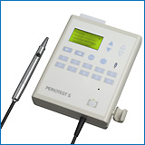
Periotest
dynamically diagnosing the human periodontium and the dental implant-bone
interface
 about
Periotest device
about
Periotest device
References: ... 19891990
19911992
19931994
19951996
19971998
19992000
2001...
Examination of the damping behavior of IMZ implants
R. HAAS, M. SABA, P.N. MENSDORFF, G. MAILATH
Int.J.Oral Maxillofac Implants 10, 410-414 (1995).
Alveolar Bone Loss diagnosis/Follow Up Studies/dental implants/Osseointegration/imz
The damping behavior of 392 IMZ implants with a diameter of 4 mm was
examined by performing Periotest measurements during regular implant
follow-up. The results were correlated with radiographically assessed peri-implant
bone reduction. To guarantee statistical independence, only one randomly
chosen implant per patient was considered in the examination. In 167 implants,
the difference between the mean mandibular and maxillary Periotest
values proved to be statistically significant. Age, sex, and radiographic
length of the endosseous part of the abutment had no influence on the Periotest
values. Assessment of the Periotest values can thus be considered
an additional parameter for objective determination of IMZ implant success.
However, exclusive use of the Periotest method without additional
radiographic examinations does not seem to be justified
Assessment of the Periotest Device in Baseline Mobility
Measurements of Craniofacial Implants
K. DERHAMI, J.F. Wolfaardt, A.Chan
Int.J.Oral Maxillofac.Impl. 10, 221-229 (1995).
Periotest/implant/Human/craniofacial/Osseointegration/mobilty
The present study aimed to evaluate reliability of the Periotest instrument
in baseline mobility measurement of craniofaeial implants in a human wet
bone specimen. Sixteen 4.0 mm craniofacial Brånemark implants were
placed in the most common sites for craniofacial implant placement. A 5.5
mm abutment was attached to each implant except one in the mastoid bone,
which was connected with a bone-anchored hearing aid (BANA) abutment. Baseline
Periotest values (PTVS) were recorded on each abutment at different vertical
measurement points by three different clinicians using three Periotest
devices. Baseline PTVs for each abutment ranged between,-I.23 and -5.90.
Interoperator and interinstrumental variability did not influence PTVS;
however, the vertical measurement point on the abutment proved to have
a strong effeet on PTV recordings.
Reprint request: Dr Kalal Derhami, Craniofacial
Osseointegration and Maxillofacial Prosthetic Rehabilitation Unit, Misericordia
Hospital, 16940-87 Avenue, Edmonton, Alberta, Canada T5R 4H5
A method for assessing the damping characteristics of periodontal
tissues: Goals and limitations
D. ROSENBERG, M. QUIRYNEN, D.V. STEENBERGHE, I.E. NAERT, J. TRICIO,
M. NYS
Quint Int 26, 191-197 (1995).
The Periotest method, an objective, noninvasive clinical diagnostic
method, is a dynamic procedure that measures the resistance of the periodontium
to a defined impact load. It has been reported that Periotest values depend
to some extent on tooth mobility, but mainly on the damping characteristics
of the periodontium. Nevertheless, the real clinical meaning of the measurements
and some important limitations of the Periotest measuring principle
still seem to be poorly understood. In the present study, the relationship
between damping characteristics of periodontal tissues and tooth mobility
was investigated. The best correlations between tooth deflection and Periotest
values were found for teeth showing a certain degree of clinical mobility
(R2 from 0.79 to 0.91). Nevertheless, this correlation was clearly lower
when only healthy subjects were examined (R2 from 0.43 to 0.54). The better
correlation found for forces greater than 1.0 N indicates that the damping
characteristics assessed with the Periotest method are related to
secondary tooth movemet. The Periotest methodology, measuring principle,
and limitations are critically reviewed.
Eine Methode zur Messung des Dämpfungsverhaltens in parodontalen
Geweben - Ziele und Einschränkungen
D. ROSENBERG, M. QUIRYNEN, D.V. STEENBERGHE, I.E. NAERT, J. TRICIO
Quintessenz 515-525 (1995).
Periodontometrie nach Mühlemann; Zahnbeweglichkeit; Zahnauslenkung
Afdeling Parodontologie, Faculteit Geneeskunde, Katholieke Universiteit,
CapuciJnenvoer 7, B-3000 Leuven:
Die Periotestmethode, ein objektives, nichtinvases klinisches Verfahreen,
ist ein dynamischer Test, der den Widerstand des Parodoritiums gegen eine
definierte Aufprallkraft prüft. Es wurde berichtet, daß die
Periotestwerte
vom Grad der Zahnbeweglichkeit, hauptsächlich jedoch vom Dämpfungsverhalten
des Parodontiurns abhängig sind. Dennoch scheinen die tatsächliche
klinische Bedeutung der Messungen und einige wichtigen Einschränkungen
des Periotest-Meßprinzips bisher unverstanden. In der vorliegenden
Studie wurde der Zusammenhang zwischen dem Dämpffungsverhalten der
parodontalen Gewebe und der Zahnbeweglichkeit untersucht. Die beste Korrelation
zwischen der Zahnauslenkung (Mühlemann-periodontometer) und den Periotestwerten
wurde bei Zähnen festgestellt, die einen gewissen Grad an klinischer
Beweglichkeit zeigten (R2 von 0,79 bis 0,91). Diese Korrolation war jedoch
deutlich geringer, wenn ausschließlich gesunde Patienten untersucht
wurden (R2 von 0,43 bis 0,54). Die bessere Korrelation erreichte man bei
einer Kraft, die größer als 1.0 N war. Dies läßt
erkennen, dass das mit der Periotestmethode gemessene Dämpfungsverhalten
mit der sekundären Zahnbeweglichkeit zusammenhängt. Außerdem
werden die Periotestmethodik, das Meßpritizip und die Einschränkungen
kritisch überprüft.
Splints made of wire and composite: an investigation of lateral
tooth mobility in vivo
K.A. EBELESEDER, K. GLOCKNER, C. PERTL, P. STÄDTLER Department
of Conservative Dentistry, Karl-Franzens-University, Graz:
Endod Dent Traumatol 11, 288-293 (1995).
Wurzelamputation; Periotest; Composite Resins; Adolescence; Child;
Linear Models; Orthodontic Wires; Percussion; Periodontal Ligament injuries;
Statistics,Nonparametric; Time Factors; Tooth Luxation complications; Tooth
Mobility etiology; Wound Healing; Periodontal Splints; Tooth Luxation therapy;
Tooth Mobility diagnosis; Tooth Mobility therapy; Female; Human; Male;
Trauma; Zahnauslenkung
In 103 posttraumatic splints, later tooth mobility was measured with
Periotest
immediately before and after the routine splint removal. The splints were
made of composite resin and an 0.017 X 0.025" orthodontic steel wire. 481
teeth were measured. A statistic evaluation revealed that the immobilisation
effect did not exceed normal tooth firmness. Fixation to one neighbouring
tooth had less effect than fixation to two. Adjacent tooth gaps reduced
the effect. Splint extensions had no influence. With the use of the Periotest
device, more than 50% of all teeth with a true mobility
of 20 Periotest-units or more were detectable as mobile in spite
of the fixed splint
Osseointegration of Branemark fixtures using a single-step operating
technique. A preliminary prospective one-year study in the edentulous mandible
BERNARD, J.P., BELSER, U.C., MARTINET, J.P., BORGIS, S.A., Department
of Oral Surgery, School of Dentistry, University of Geneva, Switzerland
Clin Oral Implants Res 6, 122-129 (1995).
Osseointegration; branemark; Mandible; implant; Adult; Aged;
Dental Plaque Index; Denture,Overlay; Feasibility Studies; Jaw,Edentulous
surgery; Mandible surgery; Middle Age; Periodontal Index; Prospective Studies;
Time Factors; Treatment Outcome; Dental Implantation,Endosseous methods;
Dental Implants
The aim of the present prospective clinical study was to analyze the
feasibility of inserting Branemark fixtures according to a one-stage procedure
including transmucosal healing and to subsequently evaluate the predictability
of osseointegration as well as the potential of such implants for stabilizing
complete overdentures in the edentulous mandible. Five patients (2 women,
3 men), completely edentulous in the mandible and with a mean age of 60
years, volunteered for this study. Two fixtures of various length (10-20
mm) and 3.75 mm in diameter were inserted in the lower canine regions.
A standard surgical procedure including a midcrestal incision was used.
After the placement of the fixtures, healing abutments, which are normally
used during second-stage surgery, were inserted instead of the usual cover
screws. Three months after implant placement a clinical and radiographic
examination was performed to confirm the presence or absence of osseointegration
of the fixtures prior to exchanging the healing abutments with the spherical
attachments. Finally, different clinical (Plaque Index, Bleeding Index,
probing depth, Periotest mobility value) and radiographic
(bone loss, peri-implant radiolucency) parameters were recorded 9 months
after loading of the fixtures by means of a complete mandibular overdenture,
retained by two ball attachments in the canine regions. All fixtures were
perfectly stable (mean Periotest values of -2) and presented favorable
peri-mplant soft tissue conditions, and no patient was complaining about
any particular symptom. As far as retention and stability of their implant
supported overdenture was concerned, the participants without exception
considered the therapeutic result as being perfectly adequate.
Influence of the suprastructure on the peri-implant tissues in beagle
dogs
HURZELER, M.B., QUINONES, C.R., SCHUPBACH, P., VLASSIS, J.M., STRUB,
J.R., CAFFESSE, R.G., Department of Stomatology, Health Science Center,
Dental Branch, University of Texas-Houston, USA
Clin Oral Implants Res 6, 139-148 (1995).
Dogs; Titanium; implant; Metals; branemark; Mandible; Acrylic
Resins; Alveolar Bone Loss etiology; Alveolar Bone Loss radiography; Analysis
of Variance; Composite Resins; Dental Plaque Index; Dental Porcelain; Dental
Prosthesis,Implant Supported adverse effects; Denture,Partial,Fixed; Metal
Ceramic Alloys; Osseointegration; Periodontal Index; Subtraction Technique;
Dental Implants adverse effects; Dental Prosthesis Design; Dental Prosthesis,Implant
Supported; Dental Implants; Omega Dental Ceramic
The purpose of this study was to compare clinical, radiographic and
histological differences around titanium oral implants loaded with either
acrylic-veneered metal or ceramo-metal fixed prostheses. Five beagle dogs
were used in this investigation. At the beginning of the study, all mandibular
premolars and first molars were extracted. After 3 months of healing, 2
Branemark implants were installed on each side of the mandibles. Three
months later, abutments were inserted on each implant and a daily oral
hygiene regime was initiated. One month after abutment connection, the
implants on one side of the mandible were restored with an acrylic-veneered
metal fixed prosthesis, whereas, on the other side a ceramo-metal fixed
prosthesis was inserted. The prostheses were constructed in occlusion with
the maxillary first molars. The following clinical parameters were measured
around each implant at this time (i.e., baseline), and thereafter, at monthly
intervals up to 5 months: Plaque Index; Gingival Index; implant mobility
(using the Periotest); probing depth and clinical attachment level
(using the Florida Probe). In addition, standardized radiographs were taken
at baseline and 5 months after insertion of the prostheses and evaluated
by subtraction radiography. Another Branemark fixture was installed on
each side of the mandibles 3 months before the end of the study. These
implants remained unloaded and submerged for the entire study period. Five
months after prosthesis insertion, the animals were killed, and implants
with their supporting peri-implant tissues were processed for histological
evaluation. Analyses of the clinical, radiographic and histometric parameters
revealed no significant differences between the acrylic-veneered and ceramometal
loaded implants.
The influence of splinting procedures on the periodontal and peri-implant
tissue damping characteristics. A longitudinal study with the Periotest
device
NAERT, I.E., ROSENBERG, D., VAN STEENBERGHE, D., TRICIO, J.A., NYS,
M., Department of Prosthetic Dentistry, Faculty of Medicine, Catholic University
of Leuven, Belgium
J Clin Periodontol 22, 703-708 (1995).
damping; Periotest; leuven; implant; Acrylic Resins; Adult; Aged;
Aged,80 and over; Biomechanics; Cementation; Denture Design; Denture,Partial,Temporary;
Follow Up Studies; Jaw,Edentulous,Partially rehabilitation; Longitudinal
Studies; Metal Ceramic Alloys; Middle Age; Monitoring,Physiologic; Osseointegration;
Dental Abutments; Dental Implantation,Endosseous; Dental Implants; Denture,Partial,Fixed;
Periodontics instrumentation; Periodontium physiology
The aim of the present study was to evaluate the capability of the
Periotest device in detecting and monitoring functional changes in the
periodontal as well as in the pari-implant damping characteristics. In
the first part of this study, 107 teeth were splinted by means of 40 full
acrylic fixed prostheses (AFP) and another 37 teeth were splinted by means
of 14 ceramometallic fixed prostheses (C-MFP). The Periotest measurements
of individual teeth were done the day the fixed prostheses were cemented
temporary (PT 1), and again after a mean observation period of 27.4 days
(PT 2). In the 2nd part, 78 osseointegrated two-stage implants were splinted
by means of 23 full acrylic fixed prosthesis (AFP) and other 18 implants
were left without it. Using the same abutment length, Periotest measurements
were performed, at abutment connections and before installation of the
final prosthesis. In a 3rd part, using both implants and teeth as abutments,
29 osseointegrated implants were connected with 25 abutment teeth by means
of 7 AFP. The measurements were performed at the beginning of the prosthetic
treatment and 2, 4 and 6 weeks later. After splinting teeth by means of
AFP for the observation period, no statistically significant reduction
in PTs was found. When on the other hand, a C-MFP was used, PT 2 showed
a significant reduction. The PTs at abutment connection went down after
a period of time, during which some implants were interconnected by means
of an AFP and others were not.
Zusammenfassung
Der Einfluß der Verblockung auf die Dämpfungseigenschaften
parodontaler und periimplantärer Gewebe. Eine Langzeitstudie mit dem
Periotest© Gerät
Mit vorliegender Studie wurde beabsichtigt, das Potential des Periotest©
Gerätes bei Entdeckung und Überwachung funktioneller Veränderungen
parodontaler, wie auch periimplantärer Dämpfungseigenschaften
zu bewerten. Im ersten Abschnitt dieser Studie wurden 107 Zähne mit
Akrylatbrücken (arcrylic fixed protheses: AFP) und weitere 37 Zähne
mit 14 Metallkeramikbrücken (ceramometallic fixed protheses: C-MFP)
verblockt. Die Periotest© Messungen der einzelnen
Zähne wurden am Tage vor der vorläufigen Zementierung (Periotestmessung
1) vorgenommen und nach einer mittleren Beobachtungszeit von 27,4 Tagen
(Periotestmessung 2) wiederholt. Im zweiten Abschnitt wurden 78
osseointegrierte zwei Phasen-Implantate mit 23 Kunststoffbrücken (AFP)
verblockt. Weitere 18 Implantate wurden unverblockt belassen. Die Periotest©
Messungen wurden anläßlich der Fixation der Pfeilerpfosten und
vor der Inkorporation der endgültigen Brücke, an Pfeilerpfosten
gleicher Länge vorgenommen. Im dritten Abschnitt wurden sowohl Implantate
als auch Zähne als Pfeiler angewandt. 29 osseointegrierte Implantate
wurden mit 25 Pfeilerzähnen durch AFP verblockt. Die Messungen wurden
zu Beginn der prothetischen Behandlung und 2,4 und 6 Wochen danach vorgenommen.
Nach AFP-Verblockung der Zähne während der Beobachtungsperiode
wurde keine wesentliche Reduktion der Periotestwerte konstatiert.
Handelte es sich dagegen um ein C-MFP, waren die Werte der Periotestmessung
2
signitfikant reduziert. Die anläßlich der Fixation der Pfeilerpfosten
gemessenen Periotestwerte verringerten sich nach einiger Zeit.
Während dieses Zeitabschnittes waren einige Implantate durch AFP verblockt
und andere nicht. Obwohl zwischen den Periotestwerten der beiden
Versorgungsvarianten kein erheblicher Unterschied vorlag, war er bei den
mit AFP verblockten Implantaten deutlicher. Auch nach einer Zeitspanne
von 6 Wochen hatte eine Verblockung von Zähnen und Implantaten in
dieser Studie zu keiner deutlichen, mit dem Periotest©
meßbaren, Veränderung der Dämpfungseigenschaften der parodontalen
Gewebe geführt.
Clinical evaluation of mandibular overdentures supported by multiple-bar
fabrication: a follow-up study of two implant systems
VERSTEEGH, P.A., VAN BEEK, G.J., SLAGTER, A.P., OTTERVANGER, J.P.,
Department of Maxillofacial Prosthodontics, De Weezenlanden Hospital, Zwolle,
The Netherlands
Int J Oral Maxillofac Implants 10, 595-603 (1995).
Follow Up Studies; implant; ITI; endosseous implants; Methods;
Alveolar Bone Loss etiology; Chi Square Distribution; Dental Implants adverse
effects; Dental Implants economics; Dental Prosthesis Retention instrumentation;
Denture,Overlay; Insurance,Dental; Middle Age; Multivariate Analysis; Osseointegration;
Periodontal Index; Proportional Hazards Models; Prosthesis Failure; Prosthesis
Related Infections etiology; Retrospective Studies; Survival Rate; Dental
Implants; Dental Prosthesis Design
A retrospective follow-up study was undertaken to assess the clinical
condition, complications, and prosthodontic aftercare of two different
implant systems over a long period. Thirty-six patients treated with a
total of 135 ITI type F endosseous implants, and 37 patients treated with
the transmandibular implants and a total of 146 transmandibular posts,
were studied during a mean follow-up period of 70 months and 44 months,
respectively. The choice of implant type was mainly influenced by a change
in financial support by the National Health Insurance Company in The Netherlands
in 1987. Cumulative success rates were calculated using the Kaplan-Meier
product limit method. In the analysis, the risk for failure of the implants
was adjusted for differences in mandibular bone height. There were no differences
between the two treatment groups with regard to age, gender, period of
edentulousness, and mandibular bone height. During the follow-up period,
plaque, bleeding, and hyperplasia scores demonstrated no significant differences
between the two groups. The ITI type F group showed significantly more
recession, and the transmandibular implant group demonstrated significantly
increased Periotest values. After adjusting for differences in bone
height, patients treated with ITI type F implants had a lower risk of failure
(relative risk, 0.55; 95% confidence interval 0.32 to 0.95). However, neither
of the implant systems fulfilled Albrektsson's criteria of success
Einflussfaktoren auf das Dämpfungsverhalten von IMZ- und Branemark-Implantaten
HAAS, R., SABA, M., MENSDORFF-POUILLY, N., SCHIEBEL, H., MAILATH, G.,
Z Zahnärztl Implantol 11, 15-18 (1995).
Periotest (R) measurements and osseointegration of mandibular ITI
implants supporting overdentures
MERICSKE-STERN, R., MILANI, D., MERICSKE, E., OLAH, A. (Bern, CH),
Clin Oral Impl Res 6, 73-82 (1995).
Die Periotest(R)-Werte von Implantaten im Unterkiefer wurden vor und
nach Belastung mit einer Hybridprothese aufgenommen und miteinander verglichen.
Es wurden 30 zahnlose Patienten (Durchschnittsalter 69 Jahre) mit 60 Bonefit-ITI-Implantaten
ausgewählt. Gemessen wurden die Periotestwert 1) nach einer Einheilphase
von 3 Monaten und 2) nachdem die Hybridprothese während 12 Monaten
getragen worden war. Beide Male wurden auch die parodontalen Parameter
aufgenommen. Zusätzlich entnahm man bei 17 Eingriffen während
der Implantation je eine Biopsie aus derselben Region des Unterkieferknochens.
Von diesen Knochen wurde die Knochendichte analysiert. Die histomorphometrische
Evaluation wurde mit der Punktzählmethode durchgeführt. Nach
Abschluss der Einheilphase lagen alle PT im Streubereich von -1 bis -8
(Mittelwert: -4.08). Ein Jahr später zeigten sich wiederum überall
negative Werte, diesmal in der Bandbreite von -2 bis -8 mit einem Mittelwert
von -4.97. Der Unterschied war statistisch signifikant (p<0.05). Die
17 Biopsien des Unterkieferknochens wurden auf ihre Dichte untersucht.
Die Knochendichte lag zwischen 22.4% und 90.9%. Man fand keine Korrelation
zwischen Knochendichte und den Periotestwert. Man konnte jedoch eine signifikante
Korrelation zwischen Ausmass der Unterkieferatrophie und der Knochendichte
feststellen
The Periotest values of mandibular implants, registered before and
after loading by overdentures, were compared. Thirty edentulous patients
with 60 Bonefit ITI implants were selected with an average age of 69 years.
The Periotest values were measured 1) after a healing period of 3 months
and 2) after the overdentures had been worn for a period of 12 months.
Periodontal parameters were recorded at both examinations. Furthermore,
17 biopsies of mandibular bone taken from the implant sites during implant
surgery were analyzed to assess the bone density. The histomorphometric
evaluation was done using a point count method. At the end of the healing
period, all registered Periotest values were negative, ranging from -1
to -8 with an average of 4.08. One year later, all measurements showed
negative values again, ranging from -2 to -8 with an average of 4.97. The
difference was statistically significant. Seventeen biopsies of mandibular
bone were evaluated to determine the density. The range of bone density
was from 22.4% and 90.9%. There was no correlation found between bone density
and Periotest values. However, a significant correlation could be observed
between mandibular atrophy and bone density. (School of Dental Medicine;
Institute of Anatomy, University of Bern, Switzerland)
Frontzahnbeweglichkeit nach direkter Klebung von Lingualretainern.
Ein Vergleich von In-vitro- und In-vivo-Messungen [The mobility
of the anterior teeth after the direct bonding of lingual retainers. A
comparison of in-vitro and in-vivo measurements]
SCHWARZE, J., BOURAUEL, C., DRESCHER, D., Poliklinik fur Kieferorthopädie,
Rheinische Friedrich- Wilhelms-Universität Bonn
Fortschr Kieferorthop 56, 25-33 (1995).
Lingual retainers are today widely used in the long-term stabilization
of incisors. In that they are left in place for a period of years, the
question arises to what extent lingual retainers allow sufficient physiological
movement. To answer this question first an in-vitro study of the inflexibility
of various retainers under horizontal and vertical load was carried out.
Next the retainer which limited physiological movement to the least degree
was studied in-vivo. Both dynamic and static mobility measurements of the
lower jaw front teeth were carried out on 5 patients before and after bonding
the retainers. A self developed measuring instrument was used to make the
static measurements and a periotest device was employed in the dynamic
measurements. The in-vitro tests indicated that, with the exception of
the retainer made of multistranded 0.0155" wire or glass fiber, a significant
reduction in tooth mobility can be expected with all the retainers. However,
the static in-vivo measurement carried out with the 0.0155" wire resulted
in a distinct reduction in tooth movement. The dynamic measurements using
the periotest device confirmed this finding. With regard to clinical practice
within the framework of long-term retention, highly flexible retainers
appear to be the most suitable and are to be recommended
Mechanical state assessment of the implant-bone continuum: a better
understanding of the Periotest method
TRICIO, J., LAOHAPAND, P., VAN STEENBERGHE, D., QUIRYNEN, M., NAERT,
I. (Department of Periodontology, Faculty of Medicine, Catholic University
of Leuven, Belgium),
Int J Oral Maxillofac Implants 10, 43-49 (1995).
The aim of this study was to obtain a better understanding of the Periotest
method when used to detect subclinical mobility of osseointegrated implants.
Four hundred two screw-shaped implants were tested with the Periotest device
at the time of abutment connection. Several factors, including jaw location,
implant and abutment length, and gender, were related to Periotest values
(PTs).
Implants located in the anterior region of the mandible showed the lowest
mean PT (- 3.2). The influence of abutment and implant length upon PTs
could be detected in the maxilla. In the mandible, only abutment length
had influence on PTs. Women showed higher mean Periotest scores in the
maxilla compared with men. This difference was not found in the mandible.
The Periotest method, its clinical limitations, and the meaning of a given
PT are also discussed
References: ... 1989 19901991
19921993
19941995
19961997
1998
19992000
2001...
Periotestwerte Tübinger Implantate
CRAMER, A., D'HOEDT, B., AXMANN, D., GOMEZ, G., SCHULTE, W.,
Gesellschaft für Orale Implantologie (Hrsg.): Jahrbuch für
Orale Implantologie, Quintessenz Verlag, Berlin (1994).
Tooth mobility and resolution of experimental periodontitis. An
experimental study in the dog
GIARGIA, M., ERICSSON, I., LINDHE, J., BERGLUNDH, T., NEIDERUD, A.M.,(Department
of Periodontology, Faculty of Odontology, University of Gothenburg, Sweden)
J Clin Periodontol 21, 457-464 (1994).
The aim of the present experiment was to study alterations in the mobility
of teeth that occurred during resolution of experimentally induced periodontitis
lesions in the dog. 5, 1- year-old, beagle dogs were used in the study.
The left and right 4th, 3rd, and 2nd mandibular premolars (4P4, 3P3, 2P2)
served as experimental teeth. Periodontal tissue breakdown was initiated
by placing plaque-collecting cotton-floss ligatures around the neck of
the experimental teeth. The ligatures were replaced to the level of the
receding gingival margin 1 x every month. On Day 120, the ligatures were
removed and debridement was performed. A groove, parallel to the long axis
of the mesial root, was prepared in the mesio-buccal surface of the crowns
of 2P and P2. Guided by the groove and with a probing force of 0.5 N, a
probe was inserted into the buccal gingival pocket of the mesial root and
was attached to the buccal surface. Biopsies including both the mesial
and distal root of 2P and P2 and the surrounding hard and soft tissues
were harvested. The biopsy procedure was repeated in a similar manner 15
days (i.e. Day 135) and 3 months (i.e. Day 225) after ligature removal
in the 4th (4P4) and 3rd (3P3) premolar regions. After fixation, decalcification
and sectioning, the biopsy material was exposed to histometric and morphometric
measurements. Assessment of the mobility of the experimental teeth was
performed on Days 120, 135 and 225 using the Periotest system.
Evaluation of the peri-implant epithelial tissue of percutaneous
implant abutments supporting maxillofacial prostheses
GITTO, C.A., PLATA, W.G., SCHAAF, N.G.,
Int J Oral Maxillofac Implants 9, 197-206 (1994).
The use of commercially pure titanium endosseous implants has become
state-of-the-art treatment for patients with craniofacial defects. This
study defined criteria that can be used in assessing the peri-implant abutment
epithelium. The criteria were then used to examine overall tissue reaction.
In this investigation, seven patients with percutaneous craniofacial implants
were evaluated. Two of these patients exhibited adverse skin reactions
that were associated with heavy sebaceous crusting, skin cultures positive
for Staphylococcus aureus, higher Periotest values, and thicker
peri-abutment tissue with greater mobility. It was determined that these
factors can predispose the patient to local infection, which, if ignored,
can result in failure of the implant. This study indicates that adequate
patient hygiene is crucial to maintaining healthy tissues in the peri-implant
abutment site
Relationship between the stiffness of the dental implant-bone system
and the duration of the implant-tapping rod contact
KANEKO, T.M., (Research Laboratory, Nikon Corporation, Tokyo, Japan)
Med Eng Phys 16, 310-315 (1994).
The physical basis of the Periotest percussion diagnosis is theoretically
studied by modelling the dental implant-bone system as a simple lumped
parameter system. An expression of the effective stiffness k(eff) is derived
as a function of the contact time tau. The validity of k(eff) obtained
is verified by a comparison with experimental values estimated from other
methods. Relationships between k(eff) and the pure stiffness for the Kelvin-Voigt
and Maxwell models are also derived. It is suggested that an increase of
viscosity as well as stiffness will decrease tau
Determination of the tooth mobility change during the orthodontic
tooth movement studied by means of Periotest and MIMD (the mechanical impedance
measuring device for the periodontal tissue)
NAKAGO, T., MITANI, S., HIJIYA, H., HATTORI, T., NAKAGAWA, Y.,(Department
of Orthodontics, Okayama University Dental School, Japan)
Am J Orthod Dentofacial Orthop 105, 92-96 (1994).
Two mechanical devices were selected to attempt to assess any changes
in tooth mobility when orthodontic force was applied. A Periotest
for assessing mobility and a mechanical impedance measuring device (MIMD)
were the instruments chosen to diagnose the changes in the periodontal
condition. The relative mobility of four canines after orthodontic tooth
movement manipulation was assessed over a 4-week period. Both devices were
able to detect very small changes in tooth mobility and could follow the
changes during tooth movement. According to the Periotest unit, the tooth
mobility changes were not within the clinically abnormal range, despite
the initial retraction load from a sectional arch, applied in a horizontal
direction with about 150 gm for each tooth. In all instances, the contiguous
maxillary first premolar had been removed 1 to 2 months before force application.
The tooth mobility observed in this investigation could be different from
that caused by periodontal disease or traumatic injury. Just after orthodontic
tooth movement manipulation, the Periotest values, as measured on the Periotest,
of tooth mobility decreased and the resistance as measured by the mechanical
impedance measuring device (MIMDM) was increased. Thus, initially, there
is a greater resistance to movement with decreased mobility. After the
experimental period of 4 weeks, there was an increase in the mobility,
as measured by the Periotest and the mechanical resistance decrements were
observed. However, the Periotest units showed some different changes
occasionally that did not seem to reflect the state of tooth movement.
This needs to be investigated further over a longer period of time. On
the basis of the results obtained, it does appear that it is feasible to
use these devices to determine mobility changes in patients at various
stages of orthodontic treatment
Beweglichkeit von Prothesenpfeilern unter dem Einfluss verschiedenartiger
Konstruktionselemente
NIEDERMEIER, W., RIESSNER, E.-M.,
Dtsch Zahnärztl Z 49, 25-29 (1994).
The influence of implant type, material, coating, diameter, and
length on periotest values at second-stage surgery: DICRG interim report
no. 4. Dental Implant Clinical Research Group
OCHI, S., MORRIS, H.F., WINKLER, S., (Department of Veterans Affairs
Medical Center, Dental Research, Ann Arbor, Michigan, USA)
Implant Dent 3, 159-162 (1994).
Many of the presently used methods of evaluating osseointegration at
second- stage surgery are highly subjective. The Periotest is claimed to
offer a more objective means to assess osseointegration by means of microcomputer-controlled
percussion. In 1991 the Dental Implant Clinical Research Group initiated
a long-term clinical study in cooperation with the Department of Veterans
Affairs to investigate the influence of implant design, application, and
site of placement on clinical performance and crestal bone height. As part
of this investigation, the Periotest values for 1, 565 root form implants
were determined at second-stage surgery and correlated with type, material,
coating, diameter, and length. Hydroxyapatite-coated implants and increased
implant diameter and length produced Periotest values that indicated a
greater extent of stability as compared with noncoated implants with shorter
diameters and lengths. Hydroxyapatite-coated cylinder-type implants yielded
the most favorable Periotest readings. Not only does the Periotest have
the potential of being a valuable instrument for assessing implant mobility
at second- stage surgery, but it also appears to have the capability of
determining slight differences in the implant-bone complex
Influence of arch bar splinting on periodontium and mobility of
fixed teeth
OIKARINEN, K.S., NIEMINEN, T.M., Department of Oral and Maxillofacial
Surgery, University of Oulu, Finland
Acta Odontol Scand 52, 203-208 (1994).
Altogether 17 patients treated with arch bar splints fixed onto teeth
were tested at the time of splint removal and approximately 5 months later.
Patients were treated with intermaxillary fixation (IMF) because of either
orthognathic surgery (7 patients) or mandibular fractures (10). The CPITN
index was used for estimating the periodontal status, and tooth mobility
was measured with Periotest. Seven patients in the orthognathic
surgery group could also be examined before splinting. Periodontal status,
as shown with relative proportions of various CPITN indexes, worsened due
to splinting but regained its original level at control examination a minimum
of 5 months after splint removal. Since the mean Periotest values did not
differ between the first and control examinations in the seven patients
undergoing orthognathic surgery, the analysis of the effect of splinting
on tooth mobility was performed from the values obtained immediately after
splint removal and at control visit. Splinting was shown to increase Periotest
values more in female patients, in younger ones, and in those who were
splinted for a shorter period. Teeth with the smallest roots showed greater
differences in Periotest values than those with large roots, and
the greatest differences in mobility were observed in incisors
Assessment of implant mobility at second-stage surgery with the
Periotest: DICRG Interim Report No. 3. Dental Implant Clinical Research
Group
TRUHLAR, R.S., MORRIS, H.F., OCHI, S., WINKLER, S., Department of Periodontics,
School of Dental Medicine, State University of New York, USA
Implant Dent 3, 153-156 (1994).
Many of the presently used methods of evaluating osseointegration at
implant uncovering are highly subjective. The Periotest is claimed to offer
a more objective means to assess osseointegration by means of microcomputer-controlled
percussion. Investigators involved in a long-term clinical study of dental
implants being conducted by the Dental Implant Clinical Research Group
used the Periotest to evaluate the mobility associated with all study implants
at second-stage surgery and correlate the Periotest values with various
bone densities. The Periotest values for 1,838 root form implants ranged
from -8 to +25. Implants that appeared to be osseointegrated at uncovering
recorded a mean Periotest value of - 3.37 +/- 3.25, while nonosseointegrated
implants had a mean Periotest value of +13.87 +/- 14.27. Mean Periotest
values were - 3.82 +/- 3.04 for quality 1 bone, -3.70 +/- 3.06 for quality
2 bone, -3.31 +/- 3.18 for quality 3 bone, and -1.29 +/- 3.57 for quality
4 bone. The Periotest has the potential of being a valuable instrument
for assessing the status of osseointegration at second-stage surgery
References: ... 1989 19901991
19921993
19941995
19961997
1998
19992000
2001...
Reconstruction of the Permaxilla with autogenous iliac bone in combination
with osseointegrated implants
APARICIO, C., BRÅNEMARK, P.-I., KELLER, E.E., OLIVÉ, J.
Minnesota, Schweden, Spanien,
Int J Oral Maxillofac Impl 8, 61-67 (1993).
Die gleichzeitige Verwendung von autologen Knochentransplantaten und
zahnärztl Implantat eröffnet neue Alternativen zur Reconstruction
von grossen Gewebedefekten. 1 Patient mit Segmentosteotomie. Beckenkammtranspl
2 spezial Brånemark Implante festsitzend Suprakonstruktion
Dämpfungsmessung an Implantatpfeilern und natürlichen
Zähnen
BUCHMANN, R., KHOURY, F., HESSLING, R., LANGE, D.E., (Münster),
Dtsch Zahnärztl Z 48, 184-187 (1993).
160 parodontal betreute Implantate und 118 natürliche Zähne
vertikale trichterförmige Knocheneinbrüche scheinen den Periotestwert
zu beeinflussen
In vitro consistency of the Periotest instrument
CHAI, J.Y., YAMADA, J., PANG, I.-C., Advanced Education Prosthodontics,
Northwestern University Dental School, Chicago, IL, USA ,J Prosthod 2,
9-12 (1993).
Consistency in measuring PT can be achieved. the consistency decreases
as mobility increases. The consistency of measuring the damping characteristics
using the Periotest (Siemens AG, Bensheim, Germany) was studied. Periotest
values (PT) of three rods embedded in various depths of impression material
were recorded by seven dentists. There is no statistically significant
difference among the subjects in measuring PT of any of the three rods.
However, the variation in the occlusal-gingival position of impact resulted
in statistically significant different PT measurements of each of the three
rods. Assessment of mobility of the same objects using a clinical criterion
was found to be less consistent as the mobility of the object increased.
In vitro PT measurements are consistent provided that the position of impact
is controlled
Assessment of oral implant mobility
CHAVEZ, H., ORTMAN, L.F., DEFRANCO, R.L., MEDIGE, J., School of Dental
Medicine, State University of New York at Buffalo, USA ,
J Prosthet Dent 70, 421-426 (1993).
load that the PT placed on the implant (eigentlich piezoelectric load
cell on stiff bench) during mobility-meas. was ~ 5 N, depending on the
compliance of the test object. srew implant (Swede-Vent entspr. Bånemark)
in vitro model embedded in blocks of self-cured aceylic resin. axial testing
machine measured the lateral displacement under a range of loads (0 to
5 N) at a crosshead speed of 0.2 mm/minute = 0.003 mm/s. A linear regression
between this stationär deviation and Periotest value
on the same models demonstrated a high level of correlation (r=0.984; b~0.01mm/PT).
Each PT unit corresponded to a deflection of approximately 0.01 mm when
a (slowly) traverse force of 5N was applied. ~enst ! in vivo with 56 Bånemark
implants. clinically successful: -6 to 02. corresponding to the in vitro
modell displacement (5N load) of 0.038 mm to 0.113 mm. These data demonstrate
that clinically successful implants are not immobile medline.
ost of the implant literature suggests that successful dental implants
are immobile and any detected mobility indicates implant failure. This
study evaluated the ability of the Periotest instrument to measure
implant mobility in a controlled in vitro model with a sample of 56 in
vivo implants. In the in vitro portion of the study, implant model mobility
was determined by an axial testing machine (0 to 5 N at 0.2 mm/minute)
and the Periotest instrument. The comparison showed a high level
of correlation (Pearson's r 0.984). The load that the Periotest
placed on the implants during mobility measurements was approximately 5
N, depending on the compliance of the test object. The in vivo portion
evaluated the range of mobility of 56 clinically successful endosseous
implants. The range of mobility as determined by the Periotest instrument
was -6 to +2. This range corresponds to an in vitro model displacement
(5 N load) of 0.038 mm to 0.113 mm with a mean of 0.066 mm +/- 0.018 mm
(SD). These data demonstrate that clinically successful implants are not
immobile, but have a range of mobility
Parodontale Parameter und deren Korrelation bei IMZ-Implantaten
im zahnlosen Unterkiefer
JANSEN, V.K., RICHTER, E.J., SPIEKERMANN, H., (Aachen),
Dtsch Zahnärztl Z 48, 207-211 (1993).
Dynamics of Periotest method of diagnosing the dental implant-bone
interface
KANEKO, T.M., (Tokyo, Japan) ,
J Mater Sc 4, 256-259 (1993).
the physical basis of the pt method has been investigated on the basis
of lumped parameter system models. theoretical values of the force to which
an implant is subjectedby tapping have been favourably compared experimental
results from literatur. 7.9N < F0 < 23N für -8
<
PT < 13.
Die Zahnbeweglichkeit lässt sich in allen Gebissphasen mit
Periotest sinnvoll und reproduzierbar bestimmen
MIETHKE, R.-R., FADEL, B.,
Prakt Kieferorthop 7, 117-128 (1993).
Die vorliegende Studie untersucht mit dem Periotestverfahren die physiologische
Beweglichkeit der Zähne im Milch-. im Wechsel- und im bleibenden Gebiss.
Das Untersuchungsgut bestand aus 80 parodontal gesunden Probanden; 39 davon
im Milchgebiss mit einem durchschnittlichen Alter von 6,0 + 0, 3
Jahren, 23 im Wechselzahngebiss mit einem Alter von 8,3 + 0,3 Jahren
und 19 im bleibenden Gebiss mit einem Durchschnittsalter von 27,0 +
6,2 Jahren. Im Wechselgebiss wurde zusätzlich die Abhängigkeit
des Periotestwertes von der Wurzellänge der Milchzähne und der
Milchmolaren im Unterkiefer überprüft. Dafür wurden die
Wurzel- und die Kronenlänge aus einem Orthopantogramm bestimmt. Dadas
Verhältnis Wurzel-/ Kronenlänge geringere Messfehler zeigte als
die absoluten Längenmasse, wurde es zur unabhängigen Variablen
der Korrelationsberechnung. Nach Einteilung der Periotestwerte in klinische
Lockerungsgrade ergibt sich für die Milchzähne der Lockerungsgrad
II (PTW 15 bis 25) im Schneidezahnbereich und der Lockerungsgrad 0 (PTW
2 bis 9) im Seitenzahnbereich beider Kiefer. Im Wechselgebiss unterscheidet
sich die physiologische Beweglichkeit der Zähne gegenüber dem
Milchgebiss nur im Schneidezahnbereich, da die bleibenden Schneidezähne
geringere Periotestwerte aufweisen ( Lockerungsgrad I (PTW 9 bis 16). Im
Schneidezahnbereich haben die männlichen Probanden in dieser Phase
höhere Periotestwerteals die weiblichen, was wahrscheinlich durch
den früheren Zahnwechsel bei Mädchen bedingt ist. Bei der röntgenologischen
Auswertung sind die geringsten Messfehler im Längenverhältnis
Wurzel-/ Kronenlänge zu finden; auberdem korreliert dieses Verhältnis
am besten mit dem Periotestwert. Lineare Korrelationskoeffizienten zwischen
Periotestwerten und Röntgenvariablen treten am deutlichsten beim 2.Milchmolaren
in Erscheinung, wo der Messfehler (aufgrund der relativ geringen Wurzelresorption)
am kleinsten ist. Die Periotestwerte der männlichen Untersuchungsgruppe
liegen hier im Durchschnitt zirka 2 unter denen der weiblichen. Dieser
geschlechtsspezifische Unterschied ist im bleibenden Gebiss am deutlichsten
zu erfassen. Insgesamt sind die Periotestwerte im Milchgebiss signifikant
höher als die Wechsel- und die im bleibenden Gebiss; das heibt, die
Zahnbeweglichkeit nimmt im Laufe der Gebissentwicklung ab. Die Oberkieferzähne
zeigen in allen drei Phasen der Gebissentwicklung höhere Periotestwerte
als diejenigen des Unterkiefers
Okklusion und parodontale Reaktion
NIEDERMEIER, W.,
Dtsch Zahnärztl Z 48, 354-361 (1993).
Failures in the osseointegration of endosseous implants
SALONEN, M.A., OIKARINEN, K., VIRTANEN, K., PERNU, H., Department of
Prosthodontics and Stomatognathic Physiology, Institute of Dentistry, University
of Oulu, Finland ,
Int J Oral Maxillofac Impl 8, 92-97 (1993).
A total of 68 patients, 26 men and 42 women, aged 21 to 86 years, were
treated with 204 endosseous implants (TPS, ITI, Bonefit, or Biolox) from
1985 to 1990. They were examined at their latest recall visit approximately
22.5 months after surgery, the range varying greatly between the implant
systems used (from 4 to 60 months). Fourteen implants were lost during
the observation period because of failures in osseointegration. There were
no statistically significant differences in success rates between the implant
systems during the observation period. The Periotest values, however,
differed between the Bonefit and ITI implants in maxillae (P < .001)
and mandibles (P < .01), and between successful and failed TPS implants
(P < .001, unpaired t test). In four of the failures, all ITI implants,
the prosthetic restorations (fixed dentures and single crowns) had been
lost. All other failures were treated by using the previous complete denture.
Possible causes of failure included advanced age and poor general health
of the patient, complications in the surgical procedures, and compromised
oral hygiene
Reproducibility and detection threshold of peri-implant diagnostics
STEENBERGHE, D.V., QUIRYNEN, M., Department of Periodontology, School
of Dentistry, Catholic University, Leuven, Belgium
Adv Dent Res 7, 191-195 (1993).
There is an increasing awareness that, for clinical monitoring of oral
implants, there is a need for reliable diagnostics and possibly prognostic
parameters. Indeed, reports have too often limited results to an inventory
of failures, while no mention was made of progressive marginal bone loss
or other symptoms of a future failure. Several parameters, such as marginal
bone level assessment and/or probing attachment level, give a precision
of up to 0.5 mm. Both measurements also seem related. The damping characteristics
of the individual implant/bone unit also offer a highly reproducible diagnostic
tool. The Periotest allows for detection of subclinical mobilities,
and 95% of repeated measurements fall within a range of one unit on the
arbitrary scale. So far, these three parameters offer no prognostic value
AD: Department of Periodontology, School of Dentistry, Catholic University,
Leuven, Belgium AB: There is an increasing awareness that, for clinical
monitoring of oral implants, there is a need for reliable diagnostics and
possibly prognostic parameters. Indeed, reports have too often limited
results to an inventory of failures, while no mention was made of progressive
marginal bone loss or other symptoms of a future failure. Several parameters,
such as marginal bone level assessment and/or probing attachment level,
give a precision of up to 0.5 mm. Both measurements also seem related.
The damping characteristics of the individual implant/bone unit also offer
a highly reproducible diagnostic tool. The Periotest allows for
detection of subclinical mobilities, and 95% of repeated measurements fall
within a range of one unit on the arbitrary scale. So far, these three
parameters offer no prognostic value
Clinical evaluation data from a comparative dental implant investigation
in dogs
STEFLIK, D.E., WHITE, S.L., PARR, G.R., SISK, A.L., SCHOEN, S.P., LAKE,
F.T., HANES, P.J., Department of Oral Pathology, Medical College of Georgia
School of Dentistry, Augusta, USA ,
J Oral Implantol 19, 199-208 (1993).
This report presents one- year clinical evaluation data from 120 ceramic
and titanium cylindrical and titanium blade-type implants placed in the
mandibles of 30 adult dogs. Ninety-six of the implants supported fixed
bridges. The bone and gingival health was evaluated by the following indices:
crevicular fluid volume index; gingival bleeding index; plaque accumulation
index; clinical mobility index; and a quantitative mobility index utilizing
the Periotest instrument. Results from this investigation suggest
that, overall, the ceramic implants exhibited more fractures and had more
mobile implants than did the titanium implant systems. Overall, complete
one-year clinical evaluation data demonstrate healthy tissue responses
to 112 of the 120 implants. Further, the Periotest instrument appears
to offer a more quantitative assessment of clinical mobility. Also, it
appears that the clinical evaluation protocol utilized in this study is
a valid procedure to use for the assessment of clinical serviceability
Reproducibility and detection threshold of peri-implant diagnostics
VAN STEENBERGHE, D., QUIRYNEN, M., Department of Periodontology, School
of Dentistry, Catholic University, Leuven, Belgium ,
Adv Dent Res 7, 191-195 (1993).
There is an increasing awareness that, for clinical monitoring of oral
implants, there is a need for reliable diagnostics and possibly prognostic
parameters. Indeed, reports have too often limited results to an inventory
of failures, while no mention was made of progressive marginal bone loss
or other symptoms of a future failure. Several parameters, such as marginal
bone level assessment and/or probing attachment level, give a precision
of up to 0.5 mm. Both measurements also seem related. The damping characteristics
of the individual implant/bone unit also offer a highly reproducible diagnostic
tool. The Periotest allows for detection of subclinical mobilities,
and 95% of repeated measurements fall within a range of one unit on the
arbitrary scale. So far, these three parameters offer no prognostic value
AD: Department of Periodontology, School of Dentistry, Catholic University,
Leuven, Belgium AB: There is an increasing awareness that, for clinical
monitoring of oral implants, there is a need for reliable diagnostics and
possibly prognostic parameters. Indeed, reports have too often limited
results to an inventory of failures, while no mention was made of progressive
marginal bone loss or other symptoms of a future failure. Several parameters,
such as marginal bone level assessment and/or probing attachment level,
give a precision of up to 0.5 mm. Both measurements also seem related.
The damping characteristics of the individual implant/bone unit also offer
a highly reproducible diagnostic tool. The Periotest allows for
detection of subclinical mobilities, and 95% of repeated measurements fall
within a range of one unit on the arbitrary scale. So far, these three
parameters offer no prognostic value
Periotest to monitor osseointegration and to check the occlusion
in oral implantology
SCHULTE, W., LUKAS, D., (Poliklinik für Zahnärztliche Chirurgie
und Parodontologie),
J Oral Implantol XIX, 23-32 (1993). call for reprints
Periotest; Osseointegration; Methods; implant; osseo integration;
occlusal
This paper reports on Periotest, its technique's history and its possible
applications. Periotest is a new diagnostic method, developed by an interdisciplinary
research group embracing dentists, physicists, mathematicians and computer
engineers. Periotest will - apart from periodontal questions -mainly assist
oral implantology
References: ... 1989 19901991
19921993
19941995
19961997
1998 19992000
2001...
YOUR COMMENTS, CRITICAL REMARKS OR SUGGESTIONS ARE VERY HELPFUL!
-
 Dieter Lukas,
update: 2002-VII-4.