Periotest
dynamically diagnosing the human periodontium and the dental implant-bone
interface
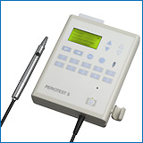 about
Periotest device
about
Periotest device
References: ... 19891990
19911992
19931994
19951996
19971998
19992000
2001...
Assessment of prosthetic restorations on bone-lock implants in patients
after oral tumor resection
A.F. KOVACS
J.Oral Implantol. 24, 101-109 (1998).
Bone Loss/Dental Implants
The feasibility of implant treatment in patients after oral ablative
tumor surgery has not yet been investigated with consideration of the requisite
high periodontal standards. A report on this topic has to deal not only
with implant survival but also with implant health, bone response, soft
tissue health, failure pattern, time of failure, and ease of restoration.
For the assessment of an implant system, an overview must be accomplished
that takes into account the different restorations used and their interaction
with the implant system that was used. This study presents the Bone-Lock
implant system (Howmedica Leibinger GmbH, Freiburg, Germany) in a retrospective
investigation after 5 years of follow-up with special emphasis on the prosthetic
restorations used following resection of oral malignancies. From early
in 1990 through June 1996, we inserted 210 dental endosteal Bone-Lock implants
(58 patients) after oral tumor resectioning. Included in the study were
45 patients with 162 implants and prosthetic restorations that had been
loaded for 1 year (dentures retained by telescopic or bar-clip or ball
attachments, implant-supported prostheses, tooth-to-implant connected bridges).
Regular follow-up consisted of evaluation of the Plaque Index (Silness
and Loe) and of the Sulcus Bleeding Index (Loe), measurements of pocket
probing depth, implant "mobility" (by means of the Periotest method),
bone resorption (according to X-ray findings), and a questionnaire that
registered patient satisfaction. The results were evaluated for each restoration
and were compared with baseline standards. The overall 5-year survival
rate was 83.2%. For implants that had been in place for over 365 days,
the survival rate was 93%. The investigation showed that after resection
of oral malignancies, patients could be treated with dental implants and
superstructures with long-term efficacy similar to that found in healthy
subjects considering internationally accepted standards. Implant treatment
in tumor patients appeared to offer the most positive periodontic results
when use of bar-clip or telescope-retained overdentures was involved. The
patient satisfaction level with the described prosthodontic treatment was
satisfactory.
Clinical and radiographic evaluation, following delivery
of fixed reconstructions, at GBR treated titanium fixtures
L. MAYFIELD, A. SKOGLUND, N. NOBREUS, R. ATTSTROM)
Clin.Oral Implants Res. 9, 292-302 (1998).
Absorbable Implants/ Alveolar Bone Loss /Dental Implants /Guided
Tissue Regeneration /Osseointegration/titanium implants
Conditions following incorporation of fixed reconstructions, at endosseous
titanium implants augmented at local bony dehiscence and fenestration defects
using a bioabsorbable Resolut membrane were studied in 7 patients. Fixture
stability, radiographic marginal bone levels and peri-implant soft tissue
status were evaluated at 21 membrane treated and 17 control fixtures (installed
in regions of adequate bone volume), following a 2-year period of functional
loading. Prosthetic reconstructions were removed and clinical examination
and Periotest values revealed that all fixtures were stable. All
peri-implant soft tissues were clinically healthy. The mean probing depths
at buccal sites for fixtures with original dehiscence (n = 10) and fenestration
(n = 11) defects were 1.6 + 0.7 mm and 1.2 + 0.4 mm respectively.
The control fixture group had a mean buccal probing depth of 1.4 +
0.6 mm. At abutment connection radiograph membrane treated fixtures had
significantly lower marginal bone levels than control fixtures, indicating
that optimal bone regeneration was not achieved at all defects. Mean radiographic
bone loss 23-27 months following delivery of fixed reconstructions for
original dehiscence and fenestration defect fixtures was 0.7 + 0.8
mm and 0.8 + 0.6 mm respectively at mesial surfaces, and 0.8 +
0.7 mm and 0.6 + 0.5 mm at distal surfaces. In the control fixture
group a mean loss of 0.7 + 0.5 mm at mesial surfaces and 0.5 +
0.4 mm at distal surfaces was found. Results showed no significant difference
in the rate of bone loss following functional loading between membrane
treated and control fixtures.
Evaluation of the Periotest as a diagnostic tool for
dental implants
A.N. CRANIN, J. DEGRADO, M. KAUFMAN, M. BARAOIDAN, R. DIGREGORIO, G.
BATGITIS, Z. LEE
J.Oral Implantol. 24, 139-146 (1998).
Alveolar Bone Loss radiography/Alveolar Bone Loss ultrasonography/Percussion
instrumentation/Periodontium radiography/Periodontium ultrasonography/dental
implants/Osseointegration/Human/Periotest
The Periotest is examined as a possible replacement for outdated,
inconsistent dental implant stability diagnosis tools. The Periotest
has the advantage of offering reproducible findings by measuring the levels
of subclinical mobility using an ultrasonically vibrating probe. The Periotest
is successful in assessing the stability status of an implant, but it can
detect the quantity of bony osseointegration only in terminal cases. Radiography
proved to be a more sensitive method of determining pericervical bone loss;
therefore, periapical radiographs in addition to the Periotest
device were found to offer the most reliable assessment of an implant's
status.
Experimental study of the damping behaviour of IMZ
implants
R. HAAS, T. BERNHART, O. DORTBUDAK, G. MAILATH
J. Oral Rehabil. 26, 19-24 (1998).
Alveolar Process anatomy and histology/Dental Prosthesis/Prognosis/Dental
Implantation/imz
Measurements of the damping behaviour of dental implants with the Periotest
device are considered to be an objective means to assess the mobility of
implants. The effects of the position of an implant in the maxilla or mandible,
the period of time passing between the measurements and implant placement
and the height at which the Periotest measurements are performed
on the damping behaviour of implants have been discussed controversially.
This experimental study examined the influence of the use of different
measuring devices, the measuring height and the embedding depth on the
damping behaviour of IMZ implants. The implants were embedded in resin
at different depths and damping measurements were carried out at different
measuring heights. It was found that the values rose with an increasing
measuring height and a decreasing embedding depth. Analysis of variance
was used to assess the influence of the embedding depth and revealed that
the embedding depth had a significant impact on the measuring values at
each measuring height, above 6 mm. Moreover, it was found that the higher
the measuring height, the higher the measured values and the greater the
differences between the values obtained at the individual depths. The different
measuring devices had no influence on the measuring results (P = 0.79).
The results of this study suggest that a longitudinal follow-up of the
peri-implant residual bone height around individual implants is possible.
Single measuring values by themselves do not allow any conclusions about
the prognosis of an implant. The assessment of the peri-implant bone height
through Periotest measurements is conceivable only when a table
of damping values taking into account the physical length of the implant,
the embedding depth and the measuring height for the examined implant system
is available. In cylindrical implants, the head of the available prefabricated
measuring post can be recommended as a constant measuring point for further
studies, especially when the results are to be compared with those obtained
by other study groups.
A comparative clinical study of three different endosseous
implants in edentulous mandibles.
A.K. ROYNESDAL, E. AMBJORNSEN, S. STOVNE, H.R. HAANAES
Int J Oral Maxillofac Implants 13, 500-505 (1998).
Bone Resorption / Dental Abutments / dental implants / Durapatite
/ Follow Up Studies / Osseointegration / Prognosis / Titanium / Treatment
Outcome / Comparative Study / Human / Prospective Studies
The purpose of this prospective study was to investigate the clinical
outcome and marginal bone resorption of three different endosseous implants
placed in the anterior mandibles of 15 elderly patients. Eleven women and
4 men (ranging from 65 to 80 years, mean 71 years) had three different
endosseous implants placed in the anterior mandible; one titanium plasma-sprayed
cylinder implant (4-mm diameter), one titanium cylinder implant with hydroxyapatite
coating (4-mm diameter), and one standard threaded titanium implant (3.75-mm
diameter). Three months later, at the second-stage surgical procedure,
ball abutments were connected and an overdenture was placed. At 12, 24,
and 36 months, marginal bone resorption and Periotest values were
recorded. None of the implants was lost in this period. An analysis of
variance with repeated measurement was performed annually to test the existence
of significant differences between the implants. When differences appeared,
paired t tests were used to identify which differences were significant.
Bonferroni multipliers were used to adjust for multiple testing. When marginal
bone resorption was concerned, threaded titanium and hydroxyapatite-coated
implants had significantly better scores than titanium plasma-sprayed cylinder
implants. Periotest values for hydroxyapatite-coated implants were
significantly better than test values for the other implants after 2 years.
After 3 years significance was obtained between hydroxyapatite and screw-shaped
implants only (P < .05). It was concluded that titanium plasma-sprayed
cylinder implants have a less favorable prognosis than the other implants
used in this study.
Untersuchungen zur Vereinfachung der Periotestmessung.
D. LUKAS, A. BÖCKLER, G. RENTSCHLER, W. SCHULTE, (Poliklinik für
Zahnärztliche Chirurgie und Parodontologie)
Dtsch Zahnärztl Z 53, 701-706 (1998)
Periotest / Percussion / mesio-exzentrisch / liegend
Zusammenfassung
Die vorliegenden Untersuchungen zum Periotestverfahren sollen klären,
ob ein Abweichen der Ausrichtung des Handstücks von der exakt orthoradialen
Perkussionsrichtung zulässig ist, da diese aufgrund anatomischer Gegebenheiten
nicht immer einzuhalten ist. Weiterhin soll festgestellt werden, ob sich
am liegenden Patienten ermittelte Periotestwerte von Messungen an sitzenden
Patienten sowohl ohne Okklusalkontakt der Zähne als auch in maximaler
Interkuspidationsposition unterscheiden.
Die Ergebnisse zeigen, dass sich sowohl für die mesialexzentrische
Messung ohne als auch mit Okklusionskontakt des Testzahnes reproduzierbare
Periotestwerte bestimmen lassen. Die Messreihenmittelwerte wiesen zum Teil
statistisch signifikante Unterschiede gegenüber den Standardmessungen
auf. Deshalb wurden Korrekturwerte erstellt, um mesialexzentrisch gemessene
Periotestwerte mit denen nach der orthoradialen Messtechnik des Periotestparodontalstatus
vergleichen zu können. Da die Korrekturwerte jedoch unterhalb der
Messgenauigkeit des Periotestgerätes von +1 Periotestwerten
für Prämolaren und +2 für Molaren lagen, kann für
Routinemessungen auf eine Korrektur verzichtet werden.
Die mesialexzentrische Perkussionsrichtung kann daher für die
Periotestmessung im Seitenzahngebiet uneingeschränkt empfohlen werden,
wodurch die Zugänglichkeit zum Testobjekt erleichtert wird. Eine unruhige
Haltung des Periotesthandstücks während der Messung muss jedoch
unbedingt vermieden werden.
Die Unterschiede der Periotestwerte von sitzenden bzw. liegenden Probanden
sind deutlich geringer als die interpersonelle Variabilität, die sich
in den Periotest-Normalbereichen manifestiert. Periotestmessungen können
somit sowohl an sitzenden als auch an liegenden Patienten durchgeführt
werden. Aus der Messung am liegenden Patienten folgt häufig eine ergonomisch
günstigere Handhabung des Periotestgerätes.
Summary
The Standard Periotest technique requires a direction of percussion
thaz is centered on the midbuccal/lingual aspect of the tooth. This technique
is not possible with every patient, due to the anatomical situation. The
possibility to use a mesial-eccentric percussing technique in molars and
premolars is investigated in this paper. Furthermore it was investigated
if Periotest values measured on patients in lying or in sitting position
are different.
The mesiallly eccentric technique just as well as percussion in midbuccal/lingual
direction was found suitable to gain reproducible Periotest values, both
without occlusal contact and in maximum intercuspidation. The mesial-eccentric
technique partially differed from standard percussion with reference to
the mean values. Because of that correction factors were calculated to
enable a comparison with the Periotest normal range which is valid with
standard technique. Since the correction factors were smaller than the
precision of the Periotest device, which is +1 with premolars and
+2
with molars, no correction is necessary in daily use.
Periotest technique with mesial-eccentric percussion in molars and
premolars is unrestrictedly recommendable, if by that handling the handpiece
is easier. Unsteadiness of the handpiece while percussing is to be avoided
absolutely.
The differences found on lying and sitting patients are only a fraction
of the interpersonal variability. Therefore Periotest measurements may
be carried out both on sitting and on lying patients. The lying position's
ergonomics is better in most cases.
Overdentures stabilised by two IMZ implants in the lower jaw--a
5-8 year retrospective study.
K. HEYDENRIJK, R.H. BATENBURG, G.M. RAGHOEBAR, H.J. MEIJER, O.R. VAN,
B. STEGENGA
Eur J Prosthodont Restor Dent 6, 19-24 (1998).
Adult / Alveolar Bone Loss / dental implants / Dental Plaque Index
/ Dental Restoration Failure / Periodontal Index / Human / IMZ
Clinical and radiographical parameters were assessed in forty patients
with overdentures stabilised by two IMZ implants connected by a bar in
the lower jaw in a 5-8 years retrospective study. Results indicated that
most patients had healthy peri-implant tissues, the mean pocket probing
depth was 3.1 mm and the median Periotest value was -4. Three implants
were removed after the healing period and replaced by three new implants.
One implant was lost after six years. One implant was mobile on palpation.
None of the patients showed objective signs of dysaesthesia in the lower
lip or chin. The peri-implant bone level of most implants had remained
stable after one year of service. The overall success rate was 94% (Albrektsson
et al.) and it is therefore concluded that two IMZ implants, connected
with a bar in the lower jaw, provide a stable base for long-term support
of a mandibular overdenture.
Reactions of peri-implant tissues to continuous loading of osseointegrated
implants.
N.N. AKIN, I. NERGIZ, A. SCHULZ, N. ARPAK, W. NIEDERMEIER
Am J Orthod Dentofacial Orthop 114, 292-298 (1998).
Bite Force / Bone Density / Compressive Strength / Densitometry,X
Ray / Dental Abutments / Dental Implants / Dogs / Periodontal Index / Alveolar
Process / Dental Stress Analysis / Orthodontic Appliances / Osseointegration
/ Animal / bonefit
Functional and morphologic reactions of peri-implant bone surrounding
screw implants (Bonefit) were studied in three dogs by loading the implants
with continuous forces of 2 (about 204 gm) and 5 N (about 510 gm). Eight
implants were inserted to an endosseous length of 12 mm and placed about
10 mm apart in the region of the lower premolars. The fixtures healed in
a closed environment for 12 weeks, after which they were uncovered and
loaded with abutments and orthodontic devices to produce horizontal distraction
with a force of 2 N (about 204 gm) for 12 weeks. Subsequently they were
loaded with 5 N (about 510 gm) for another 24 weeks. The distance between
and the mobility of the implants were determined before and after each
phase of experimental loading. Fixtures of the same type that were osseointegrated
and not exposed, or osseointegrated and loaded by mastication, were used
as a control. Animals were euthanized and specimens sectioned. The result
was that continuously loaded implants showed no significant displacement
for any force level. The mobility of the fixtures increased slightly by
about 1 Periotest-value at the end of the experiment. No significant
peri-implant pocket could be seen in implants loaded by continuous or masticatory
forces. Histologic and morphometric evaluation indicated compaction of
bone as a result of loading. Osseointegrated implants have potential as
a firm osseous anchorage for orthodontic treatment and can resist continuous
horizontal forces of at least 5 N (about 510 gm) during a period of several
months.
Die Verwendung von nicht resorbierbaren ePTFE-Membranen zur Augmentation
in der zahnärztlichen Implantologie - Ein Erfahrungsbericht.
HANDTMANN, S. (Klinik u. Poliklinik für Mund-, Kiefer- u. Gesichtschirurgie),
GÓMEZ-ROMÁN, G., AXMANN-KRCMAR, D. (Poliklinik für
Zahnärztliche Prothetik und Propädeutik),
DONATH, K. (Abteilung Oralpathologie, Institut für Pathologie
der Universität Hamburg),
LUKAS , D. (Klinik u. Poliklinik für Mund-, Kiefer- u. Gesichtschirurgie)
Quintess 49:561-575, (1998) call for reprints
Implantologie, nicht resorbierbare alloplastische Membranen, gesteuerten
Geweberegeneration (GTR), gesteuerte Knochenregeneration (GBR), Verlaufskontrollen
Die Voraussetzung für eine erfolgreiche Implantation ist das Vorliegen
eines ausreichenden Knochenvolumens sowohl in vertikaler als auch in transversaler
Richtung. Mit Hilfe der gesteuerten Geweberegeneration (GTR) unter Anwendung
von nicht resorbierbaren alloplastischen Membranen (z. B. ePTFE-Membranen)
ist es möglich, die Regeneration atrophierter Kieferbereiche zu erreichen
und damit das Implantatbett zu verbessern. Seit 1991 wird dieses operative
Verfahren auch in unserer Klinik angewandt. Insgesamt wurden mit diese
Methode bisher bei 58 Patienten mit ungünstigen anatomischen Verhältnissen
69 Implantate inseriert. Bei 50 Implantaten war der Heilungsverlauf unauffällig,
während 19 Implantate eine Exposition der Membran zeigten. 9 dieser
Membranen mußten vorzeitig entfernt werden. Insgesamt gingen 3 Implantate
vor der prothetischen Versorgung verloren. Die restlichen 66 Implantate
sind in der Zwischenzeit prothetisch versorgt und in situ. Ziel dieser
Untersuchung ist unsere Erfahrungen mit der Methode der gesteuerten Geweberegeneration
mit nicht resorbierbaren Membranen darzustellen. Bei den Jahreskontrollen
verringern sich die Periotestwerte zwischen 0 und -2 gegenüber
den Ausgangswerten bei der prothetischen Versorgung und zeigen damit eine
geringe Tendenz zur verbesserten Osseointegration.
Mobility assessment with the Periotest system in relation to histologic
findings of oral implants.
F. ISIDOR
Int J Oral Maxillofac Implants 13, 377-383 (1998).
Alveolar Bone Loss /Dental Implants /Animal/Comparative Study
The relationship between mobility assessment with the Periotest
system and histologic findings was evaluated for oral implants. Five screw-type
implants of pure titanium were placed in the mandibles of four monkeys.
Two implants in each monkey were occlusally overloaded. These
implants were brushed once a week. Plaque was allowed to accumulate around
unloaded implants with abutments in the same monkeys. During the experiment,
six of eight implants with occlusal overload showed increased manually
detectable mobility. Two of these were lost. After 18 months of experimentation,
the mobility was assessed using the Periotest system. Sections of
the implants and surrounding tissue were cut. For the excessive occlusally
loaded implants with manually detectable mobility, positive Periotest values
were recorded, and for all other implants the values were negative (range
= -7 to -2). All implants with plaque accumulation were histologically
osseointegrated but showed marginal bone loss. Two of the implants with
occlusal overload had lost osseointegration completely, and two other implants
were osseointegrated in the apical part only. A statistically significant
association between the Periotest values and the histologic bone
level or the proportion of bone-implant contact was observed. If only
clinically stable implants (i.e., without manually detectable mobility
or with a negative Periotest value) were included in the analysis,
no significant correlation was found. The Periotest values revealed
only slightly more information concerning the osseointegration of implants
than manual mobility assessments.
Summary: The main advantage of using the Periotest system,
compaired to assessing mobility manually at implants where osseointegration
already has been achieved, seems to be the reassurance of recording an
objective score, especially at implants with a questionable manually detectable
mobility.
Schlußfolgerung: Der Hauptvorteil beim Gebrauch des
Periotestgeräts - im Vergleich zur manuellen Beweglichkeitsprüfung
bei Implantaten, die erfolgreich knöchern eingeheilt sind - scheint
die Bestätigung eines objektiven Ergebnisses zu sein, besonders bei
Implantaten mit zweifelhaft feststellbarer manueller Beweglichkeit.
Periotest method: Implant-supported framework fit evaluation in
vivo
K.B. MAY, B.R. LANG, B.E. LANG, R.F. WANG
Journal of Prosthetic Dentistry 79, 648-657 (1998).
Periotest/implant/laser/Regression Analysis/Analysis of Variance
Statement of problem. In implant prosthodontics an accurate
fit of the framework to the supporting implants is paramount. However,
microgaps occur, unknown to the clinician until complications arise that
implicate errors in fit. Therefore prosthodontics would welcome a tool
or instrument that provides an objective evaluation of the fit at the implant
prosthodontic interface.Purpose. This clinical investigation determined
whether a correlation existed between the laboratory laser measurement
of the abutment analog-framework fit and the intraoral abutment-framework
fit as measured by the Periotest method.
Material and methods. Fifteen subjects received implant-supported
remote fixed partial denture supported by five (11 subjects) or six (4
subjects) implants in the mandibular jaw opposed by a complete maxillary
denture. Laser videography was used to quantify the fit of the framework
to its respective master cast with six measurements, while the fit of the
framework in the mouth was quantified with the: Periotest method.
Data were statistically analyzed with correlation analyses and multiple
regression.
Results. The overall correlation coefficient between the two
methods was r = 0.51. Regression analysis of variance revealed that the
intercept of the laser videography measurement was significant (P less
than or equal to 0.05). The mean Periotest values and standard deviation
for the abutment-framework interface were negative (-7.3 +/- 1.2). The
variance in part for the Periotest values was explained by the misfits
in the vertical axis (Delta Z, + 0.471) and in the misfit direction of
the centroids in the x-y plane (X-YVD, -0.244).
Conclusion. There was no single variable among the six measurement
variables that strongly correlated with the Periotest method in
the identification of misfit at the bearing surface as indicated by the
Periotest
value measurements. The misfit laser variables that were weakly correlated
to the Periotest values should be observable clinically with greater scrutiny.
Relationship between contact time measurements and Periotest values
when using the Periotest to measure implant stability
N. MEREDITH, B. FRIBERG, L. SENNERBY, C. APARICIO
International Journal of Prosthodontics11, 269-275 (1998).
Periotest/implant/Osseointegration/Maxilla/damping
Purpose: The Periotest is an electronic instrument that has
been advocated for the measurement of implant stability and osseointegration.
The aims of this investigation were to establish the relationship between
contact time and Periotest values when the Periotest was used to assess
implants in vivo and in vitro, and to investigate the influence of the
striking height of the Periotest handpiece and the length of implant abutment.
Materials and Methods: The accelerometer signal from a Periotest
was captured and compared with the resulting Periotest value. In vitro
measurements of Periotest values and contact time were performed on a 3-mm
abutment that had been attached to a 15-mm implant luted into an aluminium
block, and were repeated on a patient in vivo. Further measurements were
made of the abutments of six implants in turn in the maxilla of the same
patient. The standard abutment lengths on the implants were 3, 4 (x 2),
5.5 (x 2), and 7 mm.
Results: The results indicated that there was a linear relationship
between contact rime and Periotest value for implants measured in vitro
and in vivo. Greater scatter of the in vivo data was attributed to test
and patient variables including striking position, distance, and damping
as a result of the presence of soft tissues. There was a linear relationship
between the Periotest value and the striking height for implant measurements
in vivo and in vitro.
Conclusion: It can be concluded that the sensitivity of the
Periotest to clinical variables including striking height and handpiece
angulation limit the application of the instrument as a clinical diagnostic
aid to measure implant stability.
A 5-year randomized clinical trial on the influence of splinted
and unsplinted oral implants in the mandibular overdenture therapy - Part
I: peri-implant outcome
I. NAERT, S. GIZANI, M. VUYLSTEKE, D. VANSTEENBERGHE
Clinical Oral Implants Research 9, 170-177 (1998).
Implant/Prospective Studies/Treatment Outcome
Thirty-six completely edentulous patients were enrolled for a 5-year
prospective study testing the treatment outcome between splinted and unsplinted
implants retaining a mandibular hinging overdenture. The patients were
randomized into 3 groups of equal size depending on the attachment system
used such as: magnets, ball attachments or bars (reference group). Only
1 implant out of the 72 had failed at the abutment stage. Not a single
implant failed during the 5-year loading period. The accumulation of plaque
was significantly higher for the Magnet than for the Ball group. Bleeding
on probing, as well as marginal bone level, attachment level and Periotest(R)
values did not statistically differ among the groups, neither at year 1
nor at year 5. However, the Periotest(R) values were significantly
lower at year 5 compared to year 1 for all groups, which indicates a higher
rigidity at the bone-implant interface. No correlation was found between
bleeding on probing and marginal bone loss. We conclude that the connection
state of 2 implants retaining a hinging overdenture did not influence the
peri-implant outcome.
A comparative clinical study of three different endosseous implants
in edentulous mandibles
A.K. ROYNESDAL, E. AMBJORNSEN, S. STOVNE, H.R. HAANAES
International Journal of Oral and Maxillofacial Implants 13, 500-505
(1998).
implant/Mandible/Prospective Studies/Titanium/Prognosis
The purpose of this prospective study was to investigate the clinical
outcome and marginal bone resorption of three different endosseous implants
placed in the anterior mandibles of 15 elderly patients. Eleven women and
4 men (ranging from 65 to 80 years, mean 71 years) had three different
endosseous implants placed in the anterior mandible; one titanium plasma-sprayed
cylinder implant (4-mm diameter), one titanium cylinder implant with hydroxyapatite
coating (4-mm diameter), and one standard threaded titanium implant (3.75-mm
diameter). Three months later, at the second-stage surgical procedure,
ball abutments were connected and an overdenture was placed. At 12, 24,
and 36 months, marginal bone resorption and Periotestvalues were
recorded. None of the implants was lost in this period. An analysis of
valiance with repeated measurement was performed annually to test the existence
of significant differences between the implants. When differences appeared,
paired t tests were used to identify which differences were significant.
Bonferroni multipliers were used to adjust for multiple testing. When marginal
bone resorption was concerned, threaded titanium and hydroxyapatite-coated
implants had significantly better scores than titanium plasma-sprayed cylinder
implants. Periotest values for hydroxyapatite-coated implants were
significantly better than test values for the other implants after 2 years.
After 3 years significance was obtained between hpdroxyapatite and screw-shaped
implants only (P <.05). It was concluded that titanium plasma-sprayed
cylinder implants have a less favorable prognosis than the other implants
used in this study.
Use of the Endopore dental implant to restore single teeth in the
maxilla: protocol and early results.
D.A. DEPORTER, R. TODESCAN, P.A. WATSON, M. PHAROAH, D. LEVY, K. NARDINI
Int J Oral Maxillofac Implants 13, 263-272 (1998).
Crowns/Dental Abutments/Dental Implantation
This report outlines the experimental, surgical, and prosthodontic
protocols for a prospective clinical trial using the Endopore dental implant
to replace single maxillary teeth. Twenty patients (10 male, 10 female)
ranging in age from 30 to 60 years each received one implant (mean length
10.1 mm), which, after an initial healing period of 4 months, was restored
with a single crown. Records collected included radiographs, Periotestmobility
measurements, supragingival Plaque Index, and an assessment of peri-implant
soft tissue health using pocket probing depths, sulcular bleeding following
probing, and probing attachment levels. Radiographs were exposed at predetermined
intervals following crown placement (1 and 6 months, and then yearly) in
a standardized procedure using a specialized filmholder that attaches to
each implant after removal of the crown. At the time of this preliminary
report, all of the 20 implants placed had been uncovered and were in function;
16 of the implants had been in function for 6 months or more, 14 had passed
1 year of function, and 3 had passed the 2-year function point. There have
been no failures to date.
Theoretical percussion force of the periotest diagnosis.
T. KANEKO
Int J Oral Maxillofac Implants 13, 97-101 (1998).
Acceleration/Compressive Strength/Osseointegration/Percussion/implant
The Periotest percussion force acting on a dental implant was estimated
by assuming a mass-spring-dashpot model of the implant-bone system constructed
on the basis of a clinical experiment. A theoretical value of about 10
N, comparable to hitherto reported experimental values, was obtained for
an osseointegrated implant of about 1 g. The percussion force would probably
be smaller for a heavier implant.
[Endosseous implant management of tumor patients with the bone lock
system. A 5-year study]
Enossale Implantatversorgung von Tumorpatienten mit dem Bone-Lock-System.
Eine 5-Jahres-Studie.
A. KOVACS
Mund Kiefer Gesichtschir 2, 20-25 (1998).
Dental Prosthesis Design/Osseointegration /Carcinoma,Squamous Cell
rehabilitation/Dental Implantation
The implantologic rehabilitation of patients after ablative oral tumour
surgery and defect reconstruction is carried out generally without strict
assessment of the successfulness of the outcome. Therefore 210 dental implants
inserted in 58 tumour patients were subjected to regular follow-up examinations
for 5 years. The Bone-Lock osseointegrated implant system (Howmedica Leibinger,
Freiburg) was used exclusively. The plaque index (Silness and Loe), the
sulcus bleeding index (Loe), the pocket probing depth, the width of the
passive peri-implant tissue, implant mobility by means of the Periotest
method and bone resorption according to X-ray findings were ascertained.
At 60-70% of measurement points a passive peri-implant tissue was created.
After the beginning of loading, specific adaptation phenomena of tumour
patients could be detected. Despite constant plaque accumulation (mean
1.79), the bleeding disposition diminished from maximal 1.83 to 0.71. Corresponding
to this finding the pocket probing depths decreased from 5.75 mm to 4.57
mm. The implant mobility (Periotest method: mean 2.25, range -3
to +8.5) showed a decrease in the first 2 years, then the values increased.
The mobility depends on the kind of supraconstruction. Ball attachments
have the lowest and implant-supported bridges have the highest values.
Peri-implant bone resorption showed 1.43 mm as a mean value of all measurements
and had a horizontal component of 73-84%. In accordance with this the vertical
bone loss was small. After an increase during the first 2 years both values
reached a steady state around 2.5 mm. The success rate for all 210 inserted
implants is 83.2%. For implants in place for over 365 days the success
rate is 93%. Prosthetic restoration in tumour patients can be achieved
with osseointegrated dental implants according to the acknowledged international
standards.
Rigidly splinted implants in the resorbed maxilla to retain a hinging
overdenture: a series of clinical reports for up to 4 years.
I. NAERT, S. GIZANI, S.D. VAN
J Prosthet Dent 79, 156-164 (1998).
Dental implants / Dental Prosthesis / Denture,Overlay / Gingival
Hyperplasia etiology / Periodontal Index / Prospective Studies / Alveolar
Bone Loss / Periodontal Splints
STATEMENT OF PROBLEM: The results of the implant overdenture treatment
in the maxilla remains inferior to those in the mandible. Different reasons
have been alluded to, such as bone quality and quantity, number of implants,
as well as the prosthesis design.
PURPOSE: To investigate the latter, a new design for the rehabilitation
of the resorbed maxillae was set up.
MATERIAL AND METHODS: Thirteen patients were selected and provided
with four endosseous maxillary implants, splinted with a rigid-cast bar.
RESULTS: After a mean loading time of 3 years, six implants were lost;
three at abutment and another three shortly after abutment connection,
resulting in a cumulative success rate of 88.6% at year 4. A mean marginal
bone loss of 0.3 mm was observed within the first year. After the first
year, the marginal bone level, the attachment level, and the Periotest
scores hardly changed. The main prosthetic complication was the frequent
need to renew or to activate the attachments. A strong improvement in patient
satisfaction was observed when compared with the old conventional denture.
CONCLUSIONS: Within the limits of this study, the outcome confirmed
that, on a medium-term base, implant-retained hinging overdentures on four
implants were promising.
ADDRESS: Department of Prosthetic Dentistry, School of Dentistry,
Oral Pathology and Maxillofacial Surgery, Faculty of Medicine, Catholic
University of Leuven, Belgium
Vergleich von Implantationen mit und ohne simultaner
Kieferkammspreizung bei Frialit-2- und Tübinger Implantaten
[bone splitting and simultaneous implantat placement compared with
implantat insertion without bone splitting]
S. HANDTMANN, G. GÓMEZ-ROMÁN, W. SCHULTE, D. LUKAS (Poliklinik
für Zahnärztliche Chirurgie und Parodontologie)
D. AXMANN-KRCMAR (.... und Medizinische Informationsverarbeitung)
Dtsch Zahnarztl Implantol 14, 21-29 (1998) call
for reprints
Kieferkammspreizung - bone splitting
Summary: In this study the results of 70 implants in 70 patients
treated with bone splitting and simultaneous implantat placement are compared
with data of a matched control group treated with implantat insertion without
bone splitting. In the experimental group 42 Tuebingen and 28 Frialit-2
implants were placed from 1986 until 1994.Follow-up controls were carried
out until December 1995. Ten implants type Tuebingen were lost in the experimental
group and another 11 in the control group within 8 years. Of the Frialit-2
implants 2 were lost in the experimental group and none in the control
group within 4 years. Intra- and postoperative complications seen in both
groups were compared. The changes in the regular clinical parameters such
as Periotest value, sulcus depth, plaque index and gingival index
are reported. Probable causes of loss are discussed.
Zusammenfassung: In dieser Arbeit werden in einer klinischen
Studie die Methoden "Kieferkammspreizung mit simultaner Implantation" bei
70 Patienten und 70 Implantaten (42 Tübinger Implantate und 28 Frialit-2-Implantate)
mit "Implantate ohne Kieferkammspreizung" verglichen. Dazu wurden Implantate
mit und ohne Kieferkammspreizung berücksichtigt, die von 1986 bis
Ende 1994 inseriert und bis zum Stichtag Dezember 1995 beobachtet wurden.
Vor Auswertungsbeginn wurden für einen Vergleich erforderliche
Selektionskriterien festgelegtbestimmten und mit ihrer Hilfe zu der Prüfgruppe
"Kieferkammspreizung mit simultaner Implantation" eine bei OP möglichst
ähnliche Kontrollgruppe "Implantationen ohne Kieferkammspreizung"
gebildet.
Neben den intraoperativen und postoperativen Komplikationen wurden
der Verlauf der regelmäßig erhobenen Kontrollbefunde wie Periotestwerte,
Mittelwert der Sulkustiefe, Plaque- und Gingiva-Index) für beide Gruppen
dargestellt und verglichen. Die Verlustursachen werden diskutiert.
References: ... 1989 19901991
19921993
19941995
19961997
19981999
20002001...
The use of the Periotest value as the initial success criteria of
an implant: 8-year report.
C. APARICIO
Int J Periodontics Restorative Dent 17, 150-161 (1997).
Dental Abutments / Dental Alloys / Dental Restoration Failure /
Longitudinal Studies / Osseointegration / Percussion / Titanium / Dental
implants / Human / branemark / Bone Remodeling
Osseointegration is a histometric process that occurs gradually over
a period of time. The load that an implant is able to bear depends upon,
among other parameters, the quality of the bone-implant contact. For 8
years the damping capacity of 1,182 Branemark implants inserted consecutively
in 315 patients was measured using the Periotest
method. The following clinical observations were made: (1) a relationship
was found between implants with a specific Periotest
value range that, at the moment of the transepithelial connection, were
considered to be clinically stable, asymptomatic, and whose radiograph
image was not radiolucent; (2) a relationship was found between clinically
nonintegrated implants with a different Periotest
value range; (3) variations in the Periotest
value were related to the type of bone in which the implant was placed;
(4) a small percentage of borderline implants with a Periotest
value between the osseointegrated and the nonosseointegrated Periotest
values was detected; (5) the percentage of secondary failures was related
to an initial Periotest value corresponding
to a borderline implant; (6) the healing time of each implant was individualized
in accordance with the successively obtained Periotest
value; (7) the load and the design of the prosthesis were individualized;
(8) early detection of failing implants before fabrication of prostheses
is possible; (9) communication was improved between the surgeon and the
prosthodontist; and (10) variations in Periotest
value were related to bone remodeling. The sensitivity of the principal
clinical test in evaluating osseointegration is discussed regarding the
moment of its application. The use of Periotest values as an initial success
criteria of an implant is proposed.
The influence of bone quality on Periotest values of endosseous
dental implants at stage II surgery.
R.S. TRUHLAR, F. LAUCIELLO, H.F. MORRIS, S. OCHI
J Oral Maxillofac Surg 55, 55-61 (1997).
Keywords: Adult/Aged/Aged,80 and over/Bone Density/Dental Prosthesis
Design/Dental Prosthesis,Implant Supported/Dental Restoration Failure/Jaw
surgery/Jaw,Edentulous pathology/Jaw,Edentulous surgery/Jaw,Edentulous,Partially
pathology/Jaw,Edentulous,Partially surgery/Mandible pathology/Mandible
surgery/Maxilla pathology/Maxilla surgery/Middle Age/Prospective Studies/Wound
Healing/Dental Implantation,Endosseous/dental implants/Jaw pathology/Osseointegration/Female/Human/Male/Periotest/implant/Germany/
Periotest values (Periotest, Siemens AG, Bensheim, Germany) were recorded
as a baseline variable at surgical uncovering in the ongoing multicenter,
prospective clinical studies of the Dental Implant Clinical Research Group,
which uses implants from the Spectra-System (Core-Vent Corporation, Las
Vegas, NV). For 2,212 osseointegrated implants, the mean Periotest value
of mandibular implants was -4.14 (anterior, -4.22; posterior, -4.06) versus
-3.24 for maxillary implants (anterior, -2.91; posterior, -3.91). Implants
in the densest bone (quality 1) had the lowest mean Periotest value (-4.13),
followed by quality 2 (-4.00), quality 3 (-3.58), and quality 4 (-2.64).
The reliability of implant-retained hinging overdentures for the
fully edentulous mandible. An up to 9-year longitudinal study.
I.E. NAERT, M. HOOGHE, M. QUIRYNEN, S.D. VAN
Clin Oral Investig 1, 119-124 (1997).
Alveolar Bone Loss etiology/Dental Implants /Dental Restoration
/Osseointegration/Dental Prosthesis/branemark
The treatment of fully edentulous mandibles by means of implant-supported
hinging overdentures has become a routine therapy, although long-term data
on the success rate of implants and prostheses are lacking. This longitudinal
study examined 207 consecutively treated patients who received, during
the past 10 years, 449 Branemark implants to retain a mandibular hinging
overdenture, mainly on a Dolder bar. Clinical parameters and standardized
radiographs taken at every recall visit with an interval of 6-12 months
were used to judge the implant rigidity in the jawbone, the marginal bone
level, and attachment level changes. The cumulative implant failure rate
at 9 years was 3%. After loading, implant loss was concentrated during
the first 12 months, with only one failure observed at 22 months. A radiographically
determined bone loss of 0.7 mm was observed during the first year. From
the second year, a mean annual bone loss of 0.05 mm occurred. The attachment
loss, calculated as the sum of probing pocket depth plus the recession,
was 0.07 mm/year and paralleled the changes in marginal bone level over
time. The rigidity of the implant-bone interface revealed an increase over
time, as shown by a decrease in Periotest values. The bar-overdenture
complications were related to relining (23%), untightening of the retention
clip (10%), and renewal of the prosthesis (7%). When magnets or ball attachments
were used, more aftercare was needed. Fracture of the antagonistic full
denture occurred in 7% of patients. The present data indicate that the
mandibular overdenture therapy on two (Branemark) implants is a very reliable
and cost-effective treatment, even in a long-term perspective for the fixed
full mandibular prosthesis and especially in elderly patients.
Periodontal parameters of osseointegrated dental implants. A 4-year
controlled follow-up study.
K. NISHIMURA, T. ITOH, K. TAKAKI, R. HOSOKAWA, T. NAITO, M. YOKOTA
Clin Oral Implants Res 8, 272-278 (1997).
Alveolar Process radiography/Dental Plaque Index/Epithelial Attachment
/Osseointegration/Dental Implantation/Periodontal Index
The aim of this study was to evaluate the periodontal parameters of
osseointegrated dental implants. The condition of the peri-implant mucosa
was assessed using periodontal parameters, i.e., of plaque index, bleeding
on probing, probing pocket depth, probing attachment level and Periotest
scores as well as a radiographic parameter, over a 4-year follow-up period.
32 non-submerged ITI dental implants, all placed in the mandible, were
studied in 12 patients who had good oral hygiene. All patients were regularly
recalled at 6-month intervals. The overall implant success rate was 100%.
None of the implants showed any signs of inflammation, radiographic bone
loss or any detectable mobility during the follow-up period. Methods similar
to those used to evaluate the natural dentition were effectively employed
to assess the clinical status of the dental implants. The diagnostic value
of these parameters could not, however, be determined from this study due
to the absence of any peri-implant tissue complications. The results indicated
that some periodontal parameters of healthy peri-implant mucosa might be
slightly different from healthy periodontal tissue.
[Primary soft tissue coverage and specific after-care
of endosseous implants in pre-irradiated orbits]
Primare Weichgewebsbedeckung und spezifische Nachsorge enossaler
Implantate in der vorbestrahlten Orbita.
S. TVETEN, T. WEISCHER, C. MOHR
Mund Kiefer Gesichtschir 1, 289-293 (1997).
Keywords: Follow Up Studies/Orbital Neoplasms surgery/Osseointegration
radiation effects/Postoperative Complications etiology/Reoperation/Wound
Healing radiation effects/Bone Screws/Eye,Artificial/Orbit radiation effects/Orbit
Evisceration methods/Orbital Neoplasms radiotherapy/Radiation Injuries
surgery/Surgical Flaps/endosseous implants/Periotest/Osseointegration/
After orbital exenteration and high- dose irradiation (60 Gy on average),
17 endosseous implants were placed periorbitally in 5 patients. No hyperbaric
oxygen therapy was performed. All implants were primarily covered with
regional or free flaps. After second-stage surgery and aesthetic rehabilitation
with an external maxillofacial prosthesis, the pocket depth and implant
stability (Periotest) were checked, an occipitonasomental radiograph
was taken and the soft tissue assessed in short recall intervals. When
the implant was uncovered, osseointegration was stable. Within a follow-up
period of 35 months, no fixation had failed. In two patients, peri-implant
inflammation (microbiologically confirmed Staphylococcus aureus) occurred,
which was clinically only determined by soft tissue oedema and rubor. The
results demonstrate primary soft tissue covering as essential for non-irritating
implant osseointegration in the irradiated orbita. In extraoral implants
the clinical estimation of the peri-implant soft tissue, including a microbiological
examination, is required for early detection of peri-implant inflammation
in order to avoid secondary implant failure. In contrast, periotest
and pocket depth are not relevant in recognizing an ensuing peri-implant
inflammation.
The influence of maxillary sinus augmentation on the success of
dental implants through second-stage surgery.
J.W. OLSON, C.D. DENT, J.T. DOMINICI, P.M. LAMBERT, J. BELLOME, J.
BICHARA, H.F. MORRIS
Implant Dent 6, 225-228 (1997).
Dental Prosthesis Design/Osseointegration/Reoperation/Dental Implantation
An interim evaluation of the status of 102 implants placed in 42 augmented
sinuses in 27 patients involved in an alternate category of the Dental
Implant Clinical Research Group's long-term, randomized, prospective clinical
investigation is presented. Autogeneic, allogeneic, and alloplastic sinus
grafts were used, individually or mixed. Through second-stage surgery,
101 implants were found to be integrated. The mean Periotest value
at uncovering was -3.36, with a standard deviation of 4.19. The results
suggest that root form implants placed in augmented sinuses are successful
through second-stage surgery, regardless of the augmentation material or
implant design used.
Mandibular overdentures anchored to single implants: a five-year
prospective study.
G. CORDIOLI, Z. MAJZOUB, S. CASTAGNA
J Prosthet Dent 78, 159-165 (1997).
Alveolar Bone Loss pathology/Alveolar Process pathology/Dental Abutments/Dental
Plaque Index/Dental Prosthesis Design/Denture Precision Attachment/Denture
Retention/Mandible pathology/Mouth physiology/Periodontal Index/Periodontal
Pocket pathology/Periodontium physiology/dental implants/Human/Periotest
STATEMENT OF PROBLEM: It is unknown what minimum number of implants
are required to satisfactorily support and retain a mandibular overdenture.
PURPOSE: The purpose of this study was to evaluate a treatment modality
by using mandibular overdentures anchored to single implants in a geriatric
patient population.
MATERIAL AND METHODS: Twenty-one patients with a mean age of 74.2 years
were treated with single implants inserted at the mandibular midline according
to the standard two-stage surgical technique. O-Ring or ball attachments
were connected to the implants and served as overdenture anchorage. Implant
success rate, improvement of oral comfort and function, condition of the
peri-implant soft tissues, Periotest
values, and the interproximal marginal bone level were evaluated for up
to 5 years after delivery of the overdentures.
RESULTS: None of the implants were lost during the follow-up period.
A mean marginal bone loss of 1.42 + 0.56 mm was noted at 60 months.
Plaque Index was consistently high around the abutments at all reevaluation
intervals but was associated with a low Gingival Index. Pocket Depth averaged
2.41 + 0.17 mm for the entire follow-up period. Remarkable improvement
of oral comfort and function were evidenced with the overdenture treatment.
CONCLUSION: Rehabilitation with mandibular overdentures anchored to
a single implant can be a therapeutic alternative for elderly patients
experiencing discomfort and functional difficulties with conventional mandibular
dentures.
A multiparametric analysis of occlusal and periodontal jaw reflex
characteristics in young adults with normal occlusion.
S. SUDA, K. MATSUGISHI, Y. SEKI, K. SAKURAI, T. SUZUKI, S. MORITA,
K. HANADA, K. HARA
J Oral Rehabil 24, 610-613 (1997).
Bite Force/Percussion instrumentation/Percussion methods/Dental
Occlusion/Periodontal Ligament physiology/Reflex physiology/Human/Percussion/Periotest
Periodontal jaw reflex, duration of percussion sounds, tooth mobility,
and time-moment analysis of occlusal contacts by the T-scan system was
recorded in nine periodontally healthy volunteers. The results showed that
(i) reflex responses to the pressure applied to the uppercentageral incisors
in the lingolabial direction varied, depending on the background jaw-clenching
force (BCF) of the same-sided first molars. The BCF levels to elicit excitatory
reflexes were 60-80 Newton, and inhibitory reflexes were clearly elicited
with a BCF of 100 N and beyond. (ii) Duration of percussion sounds via
an occlusal sound analyser (4.73-4.84 mS: upper first molars, 4.89-5.00
mS: uppercentageral incisors) and tooth mobility using
a 'Periotest' (3.3-3.5: upper first
molars, 5.5-5.6: uppercentageral incisors) showed a normal value. (iii)
The time moments of occlusal contacts were symmetrical toward the midsagittal
axis of the occlusal plane. The centre of the anteroposterior occlusal
contacts was located in the first molar regions.
Periotest values of dental implants in the first 2 years after second-stage
surgery: DICRG interim report no. 8. Dental Implant Clinical Research
Group.
L. WALKER, H.F. MORRIS, S. OCHI
Implant Dent 6, 207-212 (1997).
Dental Implantation/Dental Prosthesis /Time Factors/Tooth Mobility
/Osseointegration
In 1991, the Dental Implant Clinical Research Group initiated a long-term
clinical study in cooperation with the Department of Veterans Affairs to
investigate the influence of implant design, application, and site of placement
on clinical performance and crestal bone height. As part of this investigation,
Periotest values for 2,212 root from implants were determined at second-stage
surgery and during a 24-month follow-up period. Mean Periotest values decreased
for implants placed in quality 1 and 2 bone, did not change for implants
in quality 3 bone, and increased for implants in quality 4 bone. Implants
in the posterior maxilla and single implants in the anterior maxilla had
increasing mean Periotest values as compared with decreasing values for
implants in other regions. Mean Periotest values for uncoated implants
decreased gradually to approach those of hydroxyapatite-coated implants.
Factors related to Periotest values in endosseal implants: a 9-year
follow-up.
M.A. SALONEN, A.M. RAUSTIA, V. KAINULAINEN, K.S. OIKARINEN (Department
of Prosthetic Dentistry and Stomatognathic Physiology, Oulu University
Hospital, Finland)
J Clin Periodontol 24, 272-277 (1997).
Alveolar Bone Loss/Bone Density/Dental Implantation/Diagnosis/Osseointegration/Periotest
Periotest values of successful endosseal implants of 2 one-stage
implant systems. TPS and ITI, were followed from 3 months to 9 years in
order to determine the factors that contribute to the values. 128 TPS screw
implants were inserted in the lower jaw of 34 subjects, (mean age 55 years),
for retaining overdentures. 108 ITI implants were inserted in the upper
and lower jaws in 50 subjects (mean age 42 years), for retaining overdentures,
crowns and bridges. Periotest values were first measured after the osseointegration
period and thereafter annually. First of all there was a difference between
the 2 implant systems. Mean Periotest values of TPS bicortical screws were
significantly lower (P < 0.05) than those of ITI implants (screws, hollow-screws,
hollow-cylinders). Factors which significantly contributed to Periotest
values of ITI implants were jaw (upper/lower), implant length and region
of the jaw in which the implant was inserted. Periotest values of ITI implants
in the lower jaw were lower than in the upper jaw (P < 0.05). The length
of implant had no effect on Periotest values in the lower jaw, but in the
upper jaw, Periotest values of ITI 8-10-mm implants were lower than 12-mm
implants (P < 0.05). Periotest value of implants inserted in the anterior
region of the upper jaw were higher than those in the posterior region
(P < 0.05). In conclusion, bone quality and implant length had a statistically
significant effect on implant mobility in long-term follow-up. Periotest
values of various implant systems, however, differ, a fact that must be
taken into account in evaluating the success of implants.
The relationship of some histologic parameters, radiographic evaluations,
and Periotest measurements of oral implants: an experimental animal study.
H. CAULIER, I. NAERT, W. KALK, J.A. JANSEN (Department of Prosthetic
Dentistry, Catholic University of Leuven, School of Dentistry, Belgium)
Int J Oral Maxillofac Implants 12, 380-386 (1997).
Bicuspid surgery/Bone Resorption etiology/Calcium Phosphates/Dental
Abutments/Dental Restoration Failure/Goats/Maxilla/Osseointegration/Percussion/Stress/Surface
Properties/Tooth Extraction/Wound Healing/Dental Implants,Single Tooth/Maxilla
surgery/Animal/Periotest/branemark
The objective of this study was to analyze the efficacy and correlation
between clinical and histologic parameters used to evaluate oral implants.
After extraction of the premolars and a healing time of 4 months in 16
Dutch goats, four Branemark implants were placed in the maxillary left
and right premolar regions. After a healing time of 6 months, followed
by another 4 months with the permucosal abutments, the goats were sacrificed
and the jaws were block-resected. Before histologic preparation, long-cone
radiographs were made and Periotest scores of the implants were recorded.
Bone level measured histomorphometrically were found to be 0.85 mm more
apically, compared to that measured radiologically (P = .001). Furthermore,
statistically significant correlations (P > 0.2) were not found between
the Periotest values of the calcium-phosphate-coated and uncoated implants
for (1) the first thread in contact with bone, or (2) with the total number
of threads in contact with bone. It was concluded that the radiologic data
overscored the real marginal bone level around screw-shaped oral implants,
and that the Periotest device is neither able to discriminate between the
first thread nor between the total number of threads in contact with bone.
A comparative study of flexible (Polyactive) versus rigid (hydroxylapatite)
permucosal dental implants. I. Clinical aspects.
G.J. MEIJER, M.S. CUNE, D.M. VAN, P.C. DE, B.C. VAN (Department of
Oral-Maxillofacial Surgery, Faculty of Medicine, University of Utrecht,
The Netherlands)
J Oral Rehabil 24, 85-92 (1997).
Bite Force/Dental Implantation,Endosseous/Dental Plaque Index/Dental
Stress /Diagnosis,Oral instrumentation/Dogs/Mandible/Materials Testing/Osseointegration/Percussion
instrumentation/Periodontal Index/Pliability/Porosity/Biocompatible Materials/dental
implants/Dental Prosthesis Design/Durapatite/Polyesters/Polyethylene Glycols/Animal/
/Polyactive/Ceramics/Titanium/Metals/Gingiva/Periotest
Recently, an elastomeric polyethyleneoxide polybutylene-terephthalate
(PEO:PBT) copolymer (Polyactive) was introduced, which exhibits bone-bonding
characteristics. In contrast to ceramics, bioglass, titanium and other
metals, PEO:PBT copolymers are flexible materials that could reduce undesirable
peak stresses along the neck of a permucosal implant. The application of
three types of Polyactive permucosal dental implants (one dense and two
porous types) and one dense hydroxylapatite (HA) implant were clinically
tested and compared during 30 weeks of loading in a dog experiment. With
respect to the porous Polyactive implants, it was observed that the pore
diameter had decreased to such an extent that optimal bone ingrowth was
not achieved. Polyactive is known to expand due to water uptake, and, as
a consequence, the sizes of the pores of the press-fit inserted implants
had decreased to a large extent. This feature explained the high loss of
the implants with the porous outer layer (6 out of 22). None of the dense
Polyactive implants and only one of the HA implants were lost. Statistical
analysis was performed solely on the dense Polyactive and the HA implants.
At the lingual sites, plaque scores, gingiva indices, bleeding indices
and corresponding pocket depths were statistically significantly lower
for both implant types, when compared to the corresponding values at the
buccal, mesial and distal sites, irrespective of the observation period.
Differences between the dense Polyactive and the HA implants were not observed.
A considerable difference in mobility was registered between the Polyactive
and the hydroxylapatite implants, as measured by the Periotest.
The dense Polyactive implants functioned adequately and had mobility resembling
natural teeth. As such, these dense flexible materials showed a favourable
clinical function and they seem promising for reducing undesired peak stresses
when applied as a dental implant.
References: ... 1989 19901991
19921993
19941995
19961997
19981999
20002001...
Comparison of Periotest values of integrated implants
with and without healing abutments: a pilot study
H.D. BARBER, R.J. SECKINGER, K. PHILLIPS, D.R. SAUNDERS, J. FORSHEY,
T. DRUMMOND, F. MANTE
Implant Dent 5, 185-187 (1996).
Periotest; implant; Osseointegration; WA; Dental Implantation,Endosseous
methods; Percussion instrumentation; Dental Abutments; Dental Implantation,Endosseous
instrumentation; dental implants; Human
The role of the Periotest in the clinical evaluation of osseointegration
has been well documented. Some clinicians have used the initial Periotest
values at second-stage surgery as a baseline to measure changes in integration
at the bone-implant interface over time. The purpose of this pilot study
was to compare Periotest values made with and without healing abutments
in place. A statistically significant difference was found between the
Periotest values that were recorded with and without healing abutments,
with the values without the healing abutments being more negative and suggestive
of greater osseointegration.
The influence of abutment surface roughness on plaque accumulation
and peri-implant mucositis.
C.M. BOLLEN, W. PAPAIOANNO, E.J. VAN, E. SCHEPERS, M. QUIRYNEN, S.D.
VAN
Clin Oral Implants Res 7, 201-211 (1996).
Bacteria isolation and purification/Ceramics chemistry/Microbial/Dental
Implantation, /Dental Plaque Index/Dental Prosthesis Design/Gingivitis
microbiology/Mouth Mucosa microbiology/Periodontal Index/Bacterial Adhesion
physiology/Dental Abutments/Dental Implants microbiology/Dental Plaque
microbiology/Gingivitis etiology /Ceramics/ Titanium
Bacterial adhesion to intra-oral, hard surfaces is firmly influenced
by the surface roughness to these structures. Previous studies showed a
remarkable higher subgingival bacterial load on rough surfaces when compared
to smooth sites. More recently, the additional effect of a further smoothening
of intra-oral hard surfaces on clinical and microbiological parameters
was examined in a short-term experiment. The results indicated that a reduction
in surface roughness below R(a) = 0.2 microns, the so-called "thresholds
R(a)", had no further effect on the quantitative/qualitative microbiological
adhesion or colonisation, neither supra- nor subgingivally. This study
aims to examine the long-term effects of smoothening intra-oral hard transgingival
surfaces. In 6 patients expecting an overdenture in the lower jaw, supported
by endosseus titanium implants, 2 different abutments (transmucosal part
of the implant): a standard machined titanium (R(a) = 0.2 microns) and
one highly polished and made of a ceramic material (R(a) = 0.06 microns)
were randomly installed. After 3 months of intra-oral exposure, supra-
and subgingival plaque samples from both abutments were compared with each
other by means of differential phase-contrast microscopy (DPCM). Clinical
periodontal parameters (probing depth, gingival recession, bleeding upon
probing and Periotest-value) were recorded around each abutment.
After 12 months, the supra- and subgingival samples were additionally cultured
in aerobic, CO2-enriched and anaerobic conditions. The same clinical parameters
as at the 3-month interval were recorded after 12 months. At 3 months,
spirochetes and motile organisms were only detected subgingivally around
the titanium abutments. After 12 months, however, both abutment-types harboured
equal proportions of spirochetes and motile organisms, both supra- and
subgingivally. The microbial culturing (month 12) failed to detect large
inter-abutment differences. The differences in number of colony- forming
units (aerobic and anaerobic) were within one division of a logarithmic
scale. The aerobic culture data showed a higher proportion of Gram-negative
organisms in the subgingival flora of the rougher abutments. From the group
of potentially "pathogenic" bacteria, only Prevotella intermedia and Fusobacterium
nucleatum were detected for anaerobic culturing and again the inter-abutment
differences were negligible. Clinically, the smoothest abutment showed
a slightly higher increase in probing depth between months 3 and 12, and
more bleeding on probing. The present results confirm the findings of our
previous short-term study, indicating that a further reduction of the surface
roughness, below a certain "threshold R(a)" (0.2 microns), has no major
impact on the supra- and subgingival microbial composition.
Correlations between radiographic, clinical and mobility parameters
after loading of oral implants with fixed partial dentures. A 2-year longitudinal
study.
U. BRAGGER, P.C. HUGEL, W. BURGIN, D. BUSER, N.P. LANG
Clin Oral Implants Res 7, 230-239 (1996).
Alveolar Bone Loss / radiography/Bone Density/Bone Remodeling/Dental
Implantation /Osseointegration/ Periodontal Attachment Loss /Periodontium
radiography/ Titanium/titanium implants/damping/Periotest
The aim of present study was to correlate the changes in the peri-implant
tissues occurring after functional loading of non-submerged titanium implants
and assessed by radiographic, clinical and mobility measurements. 11 patients
with distal extension situations received 18 implants of the ITI Dental
Implant System. After a healing period of 3 months, the suprastructures
were fabricated and seated 5 months post-surgically. For the assessment
of peri-implant bone changes, standardized vertical bitewing radiographs
with reproducible exposure geometry were evaluated using computer assisted
densitometric image analyses (CADIA) and bone height measurements. Since
the radiographic evaluations were performed at mesial and distal sites
only, the clinical parameters from these implant aspects were included
in the analysis. Clinical periodontal parameters modified for the use around
implants were obtained, damping characteristics were expressed as Periotest
readings and standardized radiographs were obtained at 1, 3, 6, 12 and
24 months after loading. In addition, radiographs were also taken at the
start of functional loading. The data obtained from this small sample of
implants demonstrated a wide range of different tissue alterations when
using radiographic, clinical and mobility assessments. The parameters of
probing attachment level (PAL) in combination with radiographic parameters
obtained at 1, 3, and 6 months after loading were good predictors for the
peri-implant tissue status at 2 years. This was shown by means of multiple
stepwise regression analyses. Mobility measurements did not reveal valuable
predictive information with the statistical models applied. Assessments
of probing attachment levels using periodontal probes rendered information
on peri-implant tissue alterations, which were closely correlated to the
radiographically measurable peri-implant bone changes.
Long-term stability of osseointegrated implants in bone regenerated
with the membrane technique. 5-year results of a prospective study with
12 implants
BUSER, K. DULA, N.P. LANG, S. NYMAN
Clin Oral Implants Res 7, 175-183 (1996).
implant; Prospective Studies; Switzerland; WA; Periotest; Prognosis;
Alveolar Bone Loss surgery; Dental Plaque Index; Follow Up Studies; Guided
Tissue Regeneration methods; Membranes,Artificial; Osseointegration; Periodontal
Index; Polytetrafluoroethylene; Alveolar Ridge Augmentation methods; Dental
Implantation,Endosseous methods; Guided Tissue Regeneration
The purpose of the present clinical study was to evaluate the 5- year
results of the first 12 implants inserted at the University of Berne in
regenerated bone following successful ridge augmentation with the membrane
technique. The patients were recalled and examined with clinical and radiographic
parameters routinely utilized in prospective studies with standard implants
in non-regenerated bone. Based on clinical and radiographic findings, all
12 implants were considered successfully integrated according to strict
criteria of success. The detailed analysis of clinical parameters revealed
no differences to results of prospective studies on standard implants in
non-regenerated bone. All implants demonstrated ankylotic stability which
was confirmed by a mean Periotest value of -2.08. The radiographic
analysis showed stable bone crest levels with a mean bone loss between
the 1- and 5-year examination of 0.30 mm. However, 2 implants exhibited
a bone loss of more than 1 mm between the 1- and 5-year examination. Therefore,
the prognosis of these 2 implants seems questionable at the present time.
It can be concluded that bone regenerated with the membrane technique reacts
to implant placement like non-regenerated bone, since all 12 implants achieved
successful tissue integration with functional ankylosis. Furthermore, this
bone is also load-bearing, since all 12 implants maintained osseointegration
over a 5-year period.
A clinical retrospective evaluation of FA/HA coated (Biocomp) dental
implants. Results after 1 year.
M.S. CUNE, R.I. VAN, P.C. DE, R.P. WILS
Clin Oral Implants Res 7, 345-353 (1996).
Dental Implants / Osseointegration/Periodontal Index/ /Titanium/Apatites/Durapatite/(TiAl6V4)/fluorapatite/Periotest
In this retrospective clinical evaluation, the performance of loaded
titanium implants with a bilayer bioactive surface coating of the relatively
reactive hydroxylapatite (HA, outer layer) and the more stable fluorapatite
(FA, inner layer) was evaluated. 98 consecutive patients were included
in the study for a total of 354 implants, loaded by overdentures, fixed
partial dentures and single tooth replacements. Statistical analyses revealed
a cumulative probability of implant survival on a per patient basis of
94.5% (s.e. 2.7%), one year after implant installation. On implants that
were lost parts of the bilayer FA/HA coating had vanished as a result of
primary or secondary infection and subsequently epithelial down growth.
Most complications were associated with implants placed in the maxilla.
Periotest
values were generally high, indicating adequate osseointegration. The Periotest
values were statistically significantly less favorable in implants placed
in the maxilla. The most favorable Periotest values and gingival
index scores were found around implants servicing overdentures. Marginal
bone loss averaged approximately 2mm during the observation period. The
amount of marginal bone loss corresponded with the distance from the neck
of the implant to the first thread of the implant's cervical screw-section.
It is concluded that, during the relatively short observation period, the
FA/HA coated implants function adequately. However, in the absence of a
direct implant-to-bone contact preceding implant loss, part of the FA/HA
coating on the titanium implants generally dissolves.
Progressive peri-implantitis. Incidence and prediction of peri-implant
attachment loss
S. JEPSEN, A. RUHLING, K. JEPSEN, B. OHLENBUSCH, H.K. ALBERS
Clin Oral Implants Res 7, 133-142 (1996).
endosseous implants; Periotest; Methods; Aged; Dental Plaque Index;
Disease Progression; Enzyme Tests; Gingival Crevicular Fluid enzymology;
Incidence; Middle Age; Observer Variation; Peptide Hydrolases metabolism;
Periodontal Attachment Loss etiology; Periodontal Attachment Loss metabolism;
Periodontal Index; Periodontics instrumentation; Periodontitis metabolism;
Predictive Value of Tests; Prognosis; Prospective Studies; Reproducibility
of Results; Sensitivity and Specificity; Statistics,Nonparametric; Dental
Implants adverse effects; Periodontal Attachment Loss diagnosis; Periodontitis
diagnosis; Periodontitis etiology; Human; Peptide Hydrolases; dental implants
The aim of this prospective study was to characterize an implant patient
population exhibiting clinical signs of peri-implantitis and to determine
subsequently the incidence of progressive attachment loss. The predictive
values of diagnostic parameters were evaluated. 25 patients with 54 endosseous
implants that had been loaded for 41 +/- 15 months were included in the
study. Clinical parameters included the assessment of plaque, bleeding
on probing, probing depth, attachment levels, and Periotest values.
Probing measurements were performed in duplicate by means of a controlled
force electronic probe (Periprobe). Peri-implant crevicular fluid samples
were collected and assayed for neutral proteolytic enzyme (NPE) activity
(Periocheck). Analysis of duplicate baseline probing data revealed a high
degree of reproducibility (mean difference: 0.1 +/- 0.3 mm). A minimum
threshold of 1.0 mm (> 3 x S.D.) loss of probing attachment was chosen
to classify a site as positive for breakdown. Alternatively, the tolerance
method was employed to identify sites with progressive attachment loss.
After 6 months, irrespective of the analytical method, 6 percent of all
sites (in 19% of the implants) and 28% of the patients had experienced
further peri-implant attachment loss. There were significant differences
(p < 0.05) in mean plaque (73% vs. 45%) and NPE (36% vs. 12%) scores
between patients with progressive peri-implantitis and those with stable
peri-implant conditions. Both bleeding on probing and the NPE-test were
characterized by high negative predictive values, and thus negative scores
can serve as indicators for stable peri-implant conditions. For monitoring
peri-implant health during recall visits, attachment level recordings with
a controlled force electronic probe in conjunction with enzymatic diagnostic
tests of the host response can be recommended .
Periodontal parameters around porous-coated dental implants after
3 to 4 years supporting overdentures
D. LEVY, D.A. DEPORTER, P.A. WATSON, R.M. PILLIAR
J Clin Periodontol 23, 517-522 (1996).
dental implants; Periotest; Analysis of Variance; Dental Implantation,Endosseous;
Dental Plaque Index; Dental Prosthesis Design; Dental Restoration Failure;
Denture,Complete,Lower; Gingival Hemorrhage pathology; Jaw,Edentulous rehabilitation;
Jaw,Edentulous surgery; Mandible; Periodontal Attachment Loss pathology;
Periodontal Pocket pathology; Porosity; Stress,Mechanical; Surface Properties;
Dental Prosthesis,Implant Supported; Denture,Overlay; Periodontal Index;
Human;
Faculty of Dentistry, University of Toronto, Canada:
In this study, an assessment using modified periodontal indices was
done on a group of 48 fully edentulous patients who had each been treated
with 3 porous-coated (EndoPore) dental implants and a mandibular overdenture.
Parameters assessed included plaque index (PI), sulcular bleeding index
(SBI), pocket probing depth (PD), probing attachment level (PAL) and mobility
(M) using a Periotest device. At the time of the assessment, all
of the patients had passed 3 years of continuous function while 26 had
passed 4 years. Approximately 50% of implant surfaces were plaque- free
while 79% of surfaces showed no bleeding upon probing. There was no correlation
between PI and SBI. The mean PD was 3.1 mm with 64% of sites < or =
3.5 mm. Mobility measurements taken with the Periotest device gave
a mean Periotest value of (-4.35) with 96% of measurements (-0.5).
No significant correlations were found between mobility and either PAL
or implant length.
A comparative study of flexible (Polyactive) versus rigid (hydroxylapatite)
permucosal dental implants. I. Clinical aspects.
Testing progressive loading of endosteal implants with the Periotest:
a pilot study.
B.E. ROTTER, R. BLACKWELL, G. DALTON
Implant Dent 5, 28-32 (1996).
Bone Density/ Percussion instrumentation/Alveolar Process/dental
implants/ Osseointegration
Notes: The progressive loading of implants was first suggested by Misch
in 1980. A major indicator of successful osseointegration is lack of implant
mobility. In a pilot study, 26 implants were placed in eight patients.
Fifteen of the implants were progressively loaded before placement of the
final restorations. The remaining 11 implants were allowed to heal with
healing abutments only and were not loaded until placement of the final
restorations. Three measurements were taken for each of the implants at
the time of uncovering and at the final restoration appointment using the
Periotest.
The mean change in Periotest values was -0.27 for the non-loaded group
as compared with -1.51 for the progressively loaded group. There was a
significantly larger increase (P < 0.001) in implant rigidity for the
progressively loaded group. Progressive loading appears to enhance implant
rigidity. The Periotest has the potential of being a valuable instrument
for the determination of osseointegration.
References: ... 1989 19901991
19921993
19941995
19961997
19981999
20002001...
YOUR COMMENTS, CRITICAL REMARKS OR SUGGESTIONS ARE VERY HELPFUL!
-
 Dieter Lukas,
update: 2002-VII-4.