Periotest
dynamically diagnosing the human periodontium and the dental implant-bone
interface
 about
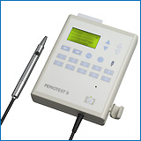
Periotest device
about
Periotest device
References: ... 1989
1990 1991
1992 1993
1994 1995
1996 1997
1998 1999
2000 2001
2002 ...
Conventional and early loading of unsplinted
ITI implants supporting mandibular overdentures.
PAYNE AG, TAWSE-SMITH A, DUNCAN WD, KUMARA R (Dunedin, New Zealand)
Clin Oral Implants 13(6):603-9 (2002)
80 and over, Alveolar Bone Loss, Cephalometry, Dental Abutments,
Dental Implants, Dental Prosthesis, Overlay, Follow-Up Studies, Human,
Middle Age, Osseointegration, Periodontal Index, Prospective Studies, Radiography,
Time Factors
The aim of this study was to compare the success rates after 1 and
2 years of conventionally and early loaded pairs of unsplinted ITI implants
supporting mandibular overdentures in edentulous patients. Twenty-four
participants (age range 55-80 years) were randomly allocated with maximum
concealment to two treatment groups. In the first group, the implants were
allowed to heal for 12 weeks before being functionally loaded (control)
and the second group had 6 weeks of healing with identical loading. All
participants had new conventional complete maxillary and mandibular dentures
prior to the study. Two sandblasted large-grit acid-etched (SLA) surface
ITI implants were placed in the mandibular interforaminal area, following
a standardized nonsubmerged surgical protocol. After 6 or 12 weeks of healing,
matrices were processed into the fitting surface of the pre-existing mandibular
dentures and the implants loaded. Implant success was determined using
mobility tests and radiographs taken at baseline and 52 and 104 weeks after
surgery. Clinical peri-implant parameters were also documented. Results
showed all implants successfully osseointegrated, according to accepted
criteria, after 2 years. Mean loss of crestal bone height after 1 year
was 0.35 +/- 0.22 mm (control) vs. 0.27 +/- 0.18 mm (test). After 2 years
this reduced to 0.09 +/- 0.06 mm (control) vs. 0.12 +/- 0.17 mm (test).
The mean Periotest value after 1 year was -4.9 (control) vs.-3.78
(test). After 2 years, the mean resonance frequency value for the control
implants was 6797 Hz [mean implant stability quotient (ISQ) = 64.77] and
for the test implants 6670 Hz (mean ISQ = 62.0). Shortened loading periods
for these ITI implants did not cause any statistically significant differences
in osseointegration or peri-implant parameters. We conclude that pairs
of unsplinted SLA-surface ITI implants can be successfully loaded with
mandibular overdentures 6 weeks after surgery.
Influence of implant geometry and surface characteristics
on progressive osseointegration.
GEURS NC, JEFFCOAT RL, MCGLUMPHY EA, REDDY MS, JEFFCOAT MK (Birmingham,
Alabama)
Int J Oral Maxillofac Implants 17(6):811-5 (2002)
Coated Materials, Biocompatible, Dental Implantation, Dental Prosthesis,
Durapatite, Human, Osseointegration, Surface Properties
PURPOSE: Although no currently available technique for the measurement
of osseointegration is entirely satisfactory, 3 clinical variables can
be reasonably associated with the process: probing depth, micromobility,
and crestal bone height. Micromobility can be quantified to some extent
with the use of the Periotest, a commercially available instrument
In this investigation, the influence of surface characteristics and geometry
upon Periotest value (PTV) and probing depth measurements was
studied. MATERIALS AND METHODS: In a multicenter trial, 120 healthy edentulous
patients received 5 or 6 implants in the anterior mandible and were followed
for 3 years. A total of 634 implants were placed. Every patient received
at least 1 implant of each of 3 types: threaded titanium plasma-sprayed
(TPS), threaded hydroxyapatite-coated (HA), and cylindric HA-coated. A
randomization schedule assured that approximately equal numbers of each
type of implant were placed and that they were uniformly distributed over
the arch. RESULTS: Of the 4 tested combinations of dependent and independent
variables, the only statistically significant (P < .05) effect was that
of coating on PTV. At 1 year after prosthetic restoration, the mean PTV
for HA-coated threaded implants was -5.36 +/- 1.24, compared with -4.86
+/- 1.70 for TPS implants. This difference steadily declined in magnitude
and significance, until, after 3 years, the groups were indistinguishable.
DISCUSSION: This study agrees with the previous observations that HA coating
tends to accelerate the initial rate of osseointegration. The absence of
a difference between threaded and cylindric implants confirms that the
PTV responds to micromobility near the surface, on a scale much smaller
than such gross geometric features. CONCLUSION: On the basis of these results,
one may conclude that HA-coated implants exhibit a more rapid decrease
in micromobility than do TPS implants of identical geometry.
Implant-retained mandibular overdentures with ITI implants
ROMEO E, CHIAPASCO M, LAZZA A, CASENTINI P, GHISOLFI M, IORIO M, VOGEL
G (Milano, Italy)
Clin Oral Implants Res 13(5):495-501 (2002)
Alveolar Bone Loss, Bone Resorption, Comparative Study, Dental Abutments,
Dental Implants, Dental Plaque Index, Dental Prosthesis, Dental Restoration
Failure, Follow-Up Studies, Gingival Hemorrhage, Human, Osseointegration,
Periodontal Pocket, Prospective Studies, Time Factors
This prospective study has been designed to compare the results of
immediate and delayed loading of implant-retained mandibular overdentures
after a 2-year follow-up. Twenty patients have been randomly divided into
two groups. Group 1 patients (test group) received four ITI implants in
the intraforaminal area of the mandible. Octa abutments were immediately
screwed on implants; 2 days after surgery, the implants were rigidly connected
with a U-shaped Dolder gold bar and loaded with an overdenture. Group 2
patients (control group) received, in the same area, the same type and
number of implants, which were left to heal according to the standard protocol.
At 3-4 months, Octa abutments were screwed on the implants and the same
prosthetic procedure of the test group was applied. The minimum follow-up
period lasted 2 years, with recall appointments at 2 weeks, 1, 3, 6 months,
1 year and every following year postoperatively, evaluating: MPI, MBI,
PD, Periotest and radiographic peri-implant bone resorption. Success
criteria according to Albrektsson et al. were used. Only one implant out
of the 40 of group 2 failed, whereas none failed in group 1. No statistical
difference of the clinical parameters evaluated was noticed in the two
groups. Therefore, immediate loading of implants, if connected with a U-shaped
bar, can provide the same results of the 'traditional' technique as far
as osseointegration and short-term survival rates of implants are concerned.
Moreover, this method significantly shortens the treatment period, thus
increasing patient satisfaction.
Clinical response to experimental forces and non-surgical
therapy of teeth with various alveolar bone loss
V. CANAKCI, R. ORBAK, A. TEZEL, C.F. CANAKCI
Dent Traumatol JID - 101091305 18, 267-274 (2002).
therapy/Alveolar Bone Loss/Periodontitis/Trauma
Firm lateral force is necessary for the thorough removal of calculus
during scaling and root planning (SRP) with hand instruments. However,
this firm lateral force should be applied to root surfaces without considering
the tooth's loss of supporting tissues. The purpose of the present study
was two-fold: firstly, to evaluate the initial pain response of periodontally
diseased non-molar teeth with two different levels of alveolar bone loss
(ABL) to experimental lateral and vertical pull forces; and secondly, to
examine the clinical response of these teeth to non-surgical therapy. Twenty
patients with chronic periodontitis were specifically selected in two groups
according to the level of ABL at non-molar teeth. Group I consisted of
10 patients who have 141 non-molar teeth with a radiographic evidence of
40-65% ABL. Group II consisted of 10 patients who have 132 non-molar teeth
with a radiographic evidence of > or =70% ABL. All patients were systemically
healthy, free of pain, and reported no current medication usage. Starting
from 0 and gradually increasing an experimental lateral force with digital
force gauge, and also an experimental vertical pull force with mechanical
force gauge were applied to each tooth and measured. As a result of a single
experimental force applied to each non-molar tooth, the initial pain response
emerged in the patients was determined by means of electronic bell system
used by patients themselves. Each patient was treated with SRP using specific
hand instruments under local anesthesia. Plaque index (PI), gingival index
(GI), probing depth (PD), clinical attachment level (CAL) and periotest
values (PV scores) were compared in both groups at initial and at month
3. A mean experimental lateral force of 24.6 N and a mean experimental
vertical pull force of 48.3 N caused initial pain response in group I.
Initial pain response occurred with a mean experimental lateral force of
5.3 N and a mean experimental vertical pull force of 19.4 N in group II.
Only group I showed statistically significant decrease in PI, GI, PD and
a significant attachment gain at month 3 (P < 0.05). There was a decrease
of 6 PV in group I at month 3 (P < 0.05), whereas an increase of 4 PV
was observed in group II (P > 0.05). This study showed that lateral and
vertical forces required for effective SRP do not cause any problem in
the group with 40-65% ABL. However, they may cause trauma in the group
with approximately 70% ABL. Thus, the results suggest that the degree of
healing would be different in the group with > or =70% ABL and in the group
with 40-65% ABL
Evaluation of implants placed with barrier membranes. A restrospective
follow-up study up to five years
M. LORENZONI, C. PERTL, R.A. POLANSKY, N. JAKSE, W.A. WEGSCHEIDER
Clin Oral Implants Res JID - 9105713 13, 274-280 (2002).
Alveolar Bone Loss/etiology/radiography/Bone Regeneration/Bone Substitutes/Bone
Transplantation/Dental Implantation,Endosseous/Follow-Up Studies/Guided
Tissue Regeneration/Membranes,Artificial
This follow-up study evaluated clinical and radiographic parameters
of dental implants placed in combination with guided bone regeneration
with barrier membranes. All implants functioned well up to 60 months after
insertion. Forty-one patients, with a total of 72 augmented implants, who
participated in a regular maintenance protocol, were investigated. Annual
Periotest
values (median value, - 3) revealed stable periimplant conditions and sustained
osseointegration. At 6 months and annually thereafter up to five years,
the radiographic evaluation yielded mean bone losses of 0.8, 1.25, 1.39,
1.42, 1.42 and 1.39 mm, respectively, with a range from 0 to 3.5 mm. No
implant failures or losses were recorded. The results demonstrated stable
periimplant conditions up to five years after membrane-protected osseous
regeneration, with no significant differences in the radiographic bone
level in regard to region, jaw or bone graft. Premature membrane exposure
resulted in a significantly higher crestal bone loss up to 24 months. The
newly formed bone appeared to be able to withstand functional loading for
up to 60 months in a predictable manner
Implant-retained mandibular overdentures with ITI implants
E. ROMEO, M. CHIAPASCO, A. LAZZA, P. CASENTINI, M. GHISOLFI, M. IORIO,
G. VOGEL
Clin Oral Implants Res JID - 9105713 13, 495-501 (2002).
overdentures/ITI implant/surgery/Bone Resorption/Osseointegration/Patient
Satisfaction
This prospective study has been designed to compare the results of
immediate and delayed loading of implant-retained mandibular overdentures
after a 2-year follow-up. Twenty patients have been randomly divided into
two groups. Group 1 patients (test group) received four ITI implants in
the intraforaminal area of the mandible. Octa(R) abutments were immediately
screwed on implants; 2 days after surgery, the implants were rigidly connected
with a U-shaped Dolder gold bar and loaded with an overdenture. Group 2
patients (control group) received, in the same area, the same type and
number of implants, which were left to heal according to the standard protocol.
At 3-4 months, Octa abutments were screwed on the implants and the same
prosthetic procedure of the test group was applied. The minimum follow-up
period lasted 2 years, with recall appointments at 2 weeks, 1, 3, 6 months,
1 year and every following year postoperatively, evaluating: MPI, MBI,
PD, Periotest(R) and radiographic peri-implant bone resorption.
Success criteria according to Albrektsson et al. were used. Only one implant
out of the 40 of group 2 failed, whereas none failed in group 1. No statistical
difference of the clinical parameters evaluated was noticed in the two
groups. Therefore, immediate loading of implants, if connected with a U-shaped
bar, can provide the same results of the 'traditional' technique as far
as osseointegration and short-term survival rates of implants are concerned.
Moreover, this method significantly shortens the treatment period, thus
increasing patient satisfaction
Stability of dental implants in microvascular osseous transplants
G. SCHULTES, A. GAGGL, H. KARCHER
Plast Reconstr Surg JID - 1306050 109, 916-921 (2002).
Dental Implantation/blood supply/transplantation/Jaw Neoplasms/surgery/Microcirculation/Reconstructive
Surgical Procedures
Microvascular iliac crest and scapula transplants have been used in
reconstruction of the lower jaw following tumor surgery. It has only been
with the insertion of dental implants that a satisfactory prosthetic rehabilitation
of the patient has been achieved. For this study, a follow-up of 38 patients
with lower jaw tumors was carried out. The patients had been treated with
partial resection of the lower jaw and neck dissection with microvascular
iliac crest transplants (n = 20) or microvascular scapula transplants (n
= 18); this was followed with dental implants (n = 143) in the region of
the transplants or the local lower jaw. One hundred thirty-nine of the
143 dental implants were loaded by prosthetic superstructures. In all patients,
the implant situation was evaluated on average 2 years 5 months after implantation.
Periotest
values, periimplant probing depths, and contact bleeding were registered,
and the extent of periimplant bone loss was defined radiographically. The
clinical situation in the region of the implant was compared for both types
of implants and also with the nonresected lower jaw. The average
Periotest
values were within the normal range for all groups. In one scapula implant,
however, a better average of Periotesting, -3.3, was found compared
with implants of the iliac crest with Periotest values of -0.7.
A measurement of -2.1 was found for the local lower jaw, similar to that
of scapula implants. Pathologic probing depths were found for all three
compared groups. The radiographically determined vertical loss of bone
was the same for all three groups, on average 1 mm at 27 months postoperatively.
The highest incidence of sulcus bleeding was found in the scapula implant
group. Thus, it can be stated that the scapula transplants provide a similar
transplant site to local lower jaw bone, whereas implants in iliac crest
transplants show lesser bony stability. Periimplant soft-tissue conditions
are worse for both types of transplants compared with local tissue of the
lower jaw
Evaluation of stability of titanium and hydroxyapatite-coated osseointegrated
dental implants: a pilot study
A. SIMUNEK, J. VOKURKOVA, D. KOPECKA, M. CELKO, R. MOUNAJJED, I. KRULICHOVA,
Z. SKRABKOVA
Clin Oral Implants Res JID - 9105713 13, 75-79 (2002).
Coated Materials,Biocompatible/Dental Alloys/dental implants/Durapatite/Elasticity/surgery/Osseointegration/Titanium/Weight-Bearing/endosseous
implants
An endosseous implant is described as osseointegrated when it is immobile
in function. Objective measures of stability testing have been described.
The Periotest is a commercially available device that is used
for this purpose. This study was designed to measure stability of endosseous
implants placed in the mandible. Implants were placed in the mandibular
canine or first premolar area to support an overdenture prosthesis. Stability
was evaluated through the use of a Periotest device at the time
of implant placement and following one year of functional loading. Implant
designs were either a screw-shaped titanium alloy or a hydroxyapatite-coated
cylinder. A total of 54 implants were placed, 37 were titanium screw-shaped
implants, while the remaining 17 were hydroxyapatite cylinders. Initial
measurements of stability showed no difference due to implant type. Following
one year of functional loading, titanium screw-shaped implants were more
stable than hydroxyapatite implants (P < 0.05). The difference in implant
rigidity following a period of functional loading may be an indication
of a difference in osseointegration between the two implants used in this
study
Porous-surfaced dental implants in the partially edentulous maxilla:
assessment for subclinical mobility
D. DEPORTER, R. TODESCAN, N. RILEY
Int J Periodontics.Restorative.Dent. 22, 184-192 (2002).
dental implants/mobility/
Abstract: Fifty patients received 151 short, porous-surfaced
implants in the partially edentulous maxilla. Periotest values
(PTV) were recorded at baseline and after 6 months and 1 and 2 years. For
this, prostheses were removed and a standard abutment attached and tightened
(20-Ncm force) to each implant. Data analysis indicated significant relationships
between time in function vs PTV and implant diameter (3.5, 4.1, or 5.0
mm) vs PTV There was no relationship between PTV and implant length. PTVs
were more favorable in the posterior than anterior maxilla, and better
PTVs were obtained with nonsplinted as opposed to splinted implants
Treatment of replacement resorption with Emdogain--a prospective
clinical study
A. FILIPPI, Y. POHL, T. VON-ARX
Dent.Traumatol. 18, 138-143 (2002).
replantation/Ankylosis/Tooth/Tooth Extraction/Trauma
The present clinical study investigated the outcome of intentional
replantation using Emdogain for periodontal healing following trauma-related
ankylosis. Sixteen ankylosed teeth affected by replacement resorption were
treated as follows: After tooth extraction, the root canal was obturated
with a retrograde titanium post. Emdogain was applied to the root surface
and into the extraction socket with subsequent replantation of the tooth.
Evaluation parameters included horizontal and vertical Periotest
scores, percussion sound and periapical radiographs. All findings were
compared to those of the adjacent teeth. The mean follow-up period was
15 months (range 4-24 months). Eleven teeth showed no signs of recurrence
of ankylosis: they were in full function and exhibited no pathological
clinical findings. Four severely traumatized teeth demonstrated a recurrence
of ankylosis after a mean period of 6 months, one tooth was lost in a second
accident after 7 months. The estimated probability of 1 year without recurrence
of ankylosis was P=0.66 (95% confidence interval [0.40; 0.94]). The mean
survival time was 10.2 months (SD 1.1). The results indicate that treatment
of replacement resorption following light to moderate trauma with replantation
and Emdogain appears to prevent or delay recurrence of ankylosis in many
cases
The transgingival approach for placement of distraction
implants
A. GAGGL, G. SCHULTES, H. RAINER, H. KARCHER
J Oral Maxillofac Surg JID - 8206428 60, 793-796 (2002).
Alveolar Bone Loss/surgery/pathology/radiography/Alveolar Ridge
Augmentation/Methods/Atrophy/Cicatrix/dental implants/Follow-Up Studies/Gingivectomy/Human/Osteotomy/Periodontitis/etiology/Periosteum/implant/complications
PURPOSE: Since 1997, distraction implants have been clinically used
for alveolar ridge distraction and, later, for prosthetic treatment. While
63 patients have been treated by the authors by alveolar ridge distraction
with distraction implants with an open approach, the aim of this study
was to demonstrate a minimally invasive technique of distractor placement
via a transgingival approach. PATIENTS AND METHODS: Twelve patients were
treated with a modified surgical incision using distraction implants. A
tissue punch was used to remove transgingival mucosa, and a segmental osteotomy
was performed using a vestibular incision. The distracted segment was pedicled
at the lingual and crestal mucoperiosteum. Distraction was carried out
for 0.5 mm per day, divided into 2 to 4 turns per day. A distraction of
5 to 7 mm was performed. At the end of distraction, the distraction insert
was changed into a definitive stable implant insert. Prosthetic treatment
was performed 4 months after the distraction period with fixed superstructures.
The follow-up was performed with the aid of dental radiographs, evaluation
of peri-implant probing depths, and Periotest values (Siemens,
Bensheim, Germany). RESULTS: The outcome of this technique showed minimal
scarring of the gingiva with good aesthetic results, the clinical and radiologic
findings were satisfying, and the Periotest values were negative
at every examination. The rate of complications was low. CONCLUSION: Minimal
scarring and good aesthetic and functional outcome resulted in patients
with alveolar ridge distraction performed with a transgingival approach.
Clinical Department of Oral and Maxillofacial Surgery, University Hospital
Graz, Auenbruggerplatz 7, A-8036 Graz, Austria alexandergaggl@kfunigrazacat
Effects of immediate loading with threaded hydroxyapatite-coated
root-form implants on single premolar replacements: a preliminary report
The transgingival approach for placement of distraction implants
P. PROUSSAEFS, J. KAN, J. LOZADA, A. KLEINMAN, A. FARNOS, A. GAGGL,
G. SCHULTES, H. RAINER, H. KARCHER
Int J Oral Maxillofac Implants 17, 567-572 (2002).
Prospective Studies/Methods/Human/surgery/mobility/Periotest/Gingiva/complications
PURPOSE: This prospective study evaluated the immediate loading of
single, threaded, root-form implants placed in the maxillary premolar area.
MATERIALS AND METHODS: Ten human subjects were included in this preliminary
report. In all cases, a screw-retained temporary acrylic resin crown was
placed immediately after implant surgery. The definitive screw-retained
metal-ceramic crown was placed 6 months later. RESULTS: Standardized radiographs
demonstrated 0.58, 0.73, 0.84, and 0.90 mm mean marginal bone loss at 1,
3, 6, and 12 months after implant surgery, respectively. Implant mobility
was evaluated with the Periotest device. At the day of surgery,
mean mobility was -3.3, while minor changes were observed thereafter: mean
values of -3.77, -3.47, and -3.63 were recorded at 3, 6, and 12 months
after implant surgery, respectively. Sulcus depth appeared relatively stable
after the 3rd month when the implant platform was used as a reference.
Recession of 0.43 mm was recorded between the 3rd and 12th month; when
the depth of the peri-implant sulcus was measured from the implant platform,
0.1 mm of change was seen between the 3rd and 12th month. Probing depth
measurements revealed that 3 months after implant placement, average probing
depth was 3.60 mm, while at 12 months it was 3.20 mm. DISCUSSION: The peri-implant
soft tissue parameters (bleeding on probing, probing depth, peri-implant
soft tissue level), mobility, and marginal bone level appeared to be similar
to findings of previous studies regarding the conventional 2-stage loading
protocol. CONCLUSION: Results of the current study provided evidence that,
under the condition of this investigation, single root-form implants can
be immediately loaded when placed in the maxillary premolar area.
Graduate Program in Implant Dentistry, Loma Linda University, California
92350, USA. pProussaef@hotmail.com
Early loading of unsplinted implants supporting mandibular
overdentures using a one-stage operative procedure with two different implant
systems: a 2-year report.
TAWSE-SMITH A, PAYNE AG, KUMARA R, THOMSON WM.Department of Oral Rehabilitation,
School of Dentistry, P.O. Box 647, University of Otago, Dunedin, New Zealand.
andrew.tawse-smith@stonebow.otago.ac.nz
Clin Implant Dent Relat Res 2002;4(1):33-42
BACKGROUND: Step-wise reduction in loading protocols is necessary to
evaluate early loading of implants with mandibular overdentures. PURPOSE:
To compare the success rates of two different dental implant systems following
conventional or early loading protocols in patients being rehabilitated
with mandibular overdentures. MATERIALS AND METHODS: Forty-eight edentulous
participants were randomly allocated to two different implant systems:
one with a machined titanium implant surface (Sterioss, Nobel Biocare,
Yorba Linda, California, USA) and the other with a roughened titanium surface
(Southern Implants, Irene, South Africa). For each system, the participants
were further divided into control groups, in whom mandibular implant overdentures
and their respective matrices were inserted following a standard 12-week
healing period, and test groups, in whom a 6-week healing period was followed
prior to identical loading. Two unsplinted implants to support implant
overdentures were placed in the anterior mandible of all participants,
using a standardized one-stage surgical procedure. Mobility tests and marginal
bone levels, as well as peri-implant parameters, were evaluated at each
baseline and 52 and 104 weeks after surgery. RESULTS: There was no statistically
significant difference in the success rates of the two systems in either
control or test groups. At the 2-year evaluation, a success rate was found
of 87.5% and 70.8% for the control and test Sterioss groups, respectively,
and 83.3% and 100% for the control and test Southern Implants groups were
observed. For the Sterioss groups, eight implants were lost at an early
stage: seven in the test group and one in the control group. For the Southern
Implants control and test groups, no failures were seen at any time interval.
There were no significant differences in marginal bone loss, Periotest
values, and peri-implant parameters between implant systems or between
any of the control or test groups. CONCLUSIONS: Early loading, with
step-wise reductions in loading protocols, of unsplinted machined Sterioss
and roughened Southern Implants fixtures with mandibular overdentures is
possible for up to 2 years.
Enhancement of primary stability of dental implants using cortical
satellite implants.
ENGELKE W, STAHR S, SCHWARZWALLER W. Department of Oral Surgery, School
of Dentistry, Georg-August-University Gottingen, Gottingen, Germany. wengelke@med.uni-goettingen.de
Implant Dent 2002;11(1):52-7
PURPOSE: This study aims to assess the effect of satellite implants
on the primary stability of dental implants placed in fresh extraction
sites in vitro. METHOD: 34 titanium screw implants (3.75 mm x 10 mm; Bego,
Bremen, Germany) were inserted in premolar- and molar-fresh extraction
sites in domestic pig mandibles. Periotest (PT) values were assessed
before and after insertion of one vestibular and one lingual 1.7-mm bone
screw (Mondeal, Tuttlingen, Germany) as a satellite implant was connected
to the implants with a 0.6-mm microplate welded to the implant abutment.
RESULTS: The average PT values were 2.9 without satellite implants, -1.0
with one satellite implant, and -2.5 with two satellite implants during
horizontal testing, and 3.0, 1.4, and 0.4, respectively, for vertical testing.
CONCLUSION: Satellite implants increase the horizontal stability of implants
in fresh extraction sites. Differences for horizontal PT assessment were
significant on a 0.01 level of confidence. Implants in extraction sites
may be loaded immediately, if vertical stabilization is provided by cortical
bone and if horizontal PT values show sufficient stability after satellite
implant insertion.
Stability of dental implants in microvascular osseous transplants.
SCHULTES G, GAGGL A, KARCHER H. Department of Oral and Maxillofacial
Surgery, University Hospital Graz.
Plast Reconstr Surg 2002 Mar;109(3):916-21
Microvascular iliac crest and scapula transplants have been used in
reconstruction of the lower jaw following tumor surgery. It has only been
with the insertion of dental implants that a satisfactory prosthetic rehabilitation
of the patient has been achieved. For this study, a follow-up of 38 patients
with lower jaw tumors was carried out. The patients had been treated with
partial resection of the lower jaw and neck dissection with microvascular
iliac crest transplants (n = 20) or microvascular scapula transplants (n
= 18); this was followed with dental implants (n = 143) in the region of
the transplants or the local lower jaw. One hundred thirty-nine of the
143 dental implants were loaded by prosthetic superstructures. In all patients,
the implant situation was evaluated on average 2 years 5 months after implantation.
Periotest
values, periimplant probing depths, and contact bleeding were registered,
and the extent of periimplant bone loss was defined radiographically. The
clinical situation in the region of the implant was compared for both types
of implants and also with the nonresected lower jaw. The average
Periotest
values were within the normal range for all groups. In one scapula implant,
however, a better average of Periotesting, minus sign3.3, was
found compared with implants of the iliac crest with
Periotest
values of minus sign0.7. A measurement of minus sign2.1 was found for the
local lower jaw, similar to that of scapula implants. Pathologic probing
depths were found for all three compared groups. The radiographically determined
vertical loss of bone was the same for all three groups, on average 1 mm
at 27 months postoperatively. The highest incidence of sulcus bleeding
was found in the scapula implant group. Thus, it can be stated that the
scapula transplants provide a similar transplant site to local lower jaw
bone, whereas implants in iliac crest transplants show lesser bony stability.
Periimplant soft-tissue conditions are worse for both types of transplants
compared with local tissue of the lower jaw.
Single-tooth replacement with the Frialit-2 system: a retrospective
clinical analysis of 146 implants
G. KRENNMAIR, S. SCHMIDINGER, O. WALDENBERGER
Int J Oral Maxillofac Implants 17, 78-85 (2002).
dental implant/Bone Resorption/
PURPOSE: This study was intended to provide a report of experience
and results with Frialit-2 implants used for single-tooth replacement.
MATERIALS AND METHODS: Over a 7-year period (1994-2000), 146 single-tooth
implants (84 maxilla, 62 mandible) were placed in 112 patients (67 females,
45 males; 31.2 +/- 16.4 years). The sites included maxillary anterior teeth
(n = 38) as well as the mandibular premolars and molars (n = 57). Ninety-three
crowns were cemented and 53 crowns were screw mounted (22 with vertical,
31 with horizontal screws) on standard abutments. The follow-up time varied
between 3 and 80 months (35.8 +/- 16.5 months). RESULTS: Two implants (1.4%)
were lost, 1 during early loading and the other after 6 years. The most
frequent prosthetic complication was isolated crown loosening of cemented
crowns requiring recementation of 9 crowns (9.9%). Crowns with vertical
screws showed no crown and/or screw loosening. Four crowns (2.8%) were
replaced because of ceramic fracture. DISCUSSION: Peri-implant soft tissue
condition, bone resorption, and Periotest values indicated satisfactory
results. The cumulative implant survival rate during the follow-up period
was 97.3%, and that of the crowns 96.4% (total cumulative survival rate
93.7%). CONCLUSIONS: With the low number of abutment screw loosenings (3.5%),
the deep internal hexagonal retention compared favorably to external retention
methods. The predominant use of long implants (98.4% > or = 13 mm) allowed
a favorable implant/crown ratio with the potential for problem-free, long-term
results
Peri-implant tissue response of immediately loaded, threaded, HA-coated
implants: 1-year results
K. RUNGCHARASSAENG, J.L. LOZADA, J.Y. KAN, J.S. KIM, W.V. CAMPAGNI,
C.A. MUNOZ
J Prosthet Dent 87, 173-181 (2002).
dental implant/overdentures/
Statement of Problem. Although high success rates have been reported
with immediately loaded implants, the peri-implant tissue response has
not been well documented. Purpose. This study evaluated implant success
and peri-implant tissue response of immediately loaded, threaded, hydroxyapatite
(HA)-coated root-form implants supporting mandibular bar overdentures with
opposing conventional maxillary complete dentures in humans. Material and
Methods. Five patients (3 men, 2 women; mean age 61 years) each received
4 HA-coated endosseous root-form implants in the interforaminal region
in the mandible. The implants were rigidly splinted with a metal framework
within 24 hours. The final EDS clip prosthesis was placed 1 to 2 weeks
thereafter. The implants and peri-implant tissues were evaluated clinically
and radiographically 0, 1, 3, 6, and 12 months after prosthesis placement.
Data were analyzed with a repeated measures 1-way analysis of variance
(P<.05). Results. All implants were stable at the end of the observation
period (mean Periotest value = minus sign5.9 plus minus 1.4).
No peri-implant radiolucencies were noted, and no implants were lost. The
mean marginal bone changes were minus sign0.42 plus minus 0.34, minus sign0.84
plus minus 0.55, minus sign1.14 plus minus 0.80, and minus sign1.16 plus
minus 0.89 mm at the 1-, 3-, 6-, and 12-month follow-ups, respectively
(P<.001). Significant declines in the rates of marginal bone changes
at each time interval were noted (P<.001). In addition, there were significant
decreases in probing depth (P<.001) and plaque index (P<.001) but
no significant difference in the frequency of bleeding upon probing (P=.64).
Conclusion. Within the limitations of this study, the peri-implant tissue
response of immediately loaded, HA-coated implants was favorable and comparable
to that of conventional, delayed-loaded implants after 1 year
Implant-retained mandibular overdentures with Branemark System MKII
implants: a prospective comparative study between delayed and immediate
loading
M. CHIAPASCO, S. ABATI, E. ROMEO, G. VOGEL
Int J Oral Maxillofac Implants 16, 537-546 (2001).
Bone Resorption/radiography/Dental Implantation/Dental Plaque Index/Dental
Prosthesis/Denture,Overlay/Osseointegration/
This study was designed to compare the results of immediate and delayed
loading of implants with implant-retained mandibular overdentures. Ten
patients (test group) received 40 Branemark System MKII implants (4 per
patient) placed in the interforaminal area of the mandible. Standard abutments
were immediately screwed to the implants, rigidly connected with a bar,
and immediately loaded with an overdenture. Ten patients (control group)
received the same type and number of implants in the same area, but the
implants were left to heal submerged. Four to 8 months later, standard
abutments were screwed to the implants and the same prosthetic procedure
was applied. Each implant was evaluated at the time of prosthetic loading
and at 6, 12, and 24 months after the initial prosthetic load with the
following parameters: modified Plaque Index (MPI), modified Bleeding Index
(MBI), probing depth (PD), and Periotest. Peri-implant bone resorption
was evaluated on panoramic radiographs taken 12 and 24 months after initial
prosthetic loading. No significant differences were found between the 2
groups regarding MPI, MBI, Periotest, peri-implant bone resorption, and
PD at 6 and 24 months (P > .05). The only difference was found regarding
PD values on the mesial and lingual sites at 12 months (P < .05). The
cumulative success rate of implants was 97.5% in both groups. Results from
this study showed that immediate loading of endosseous implants rigidly
connected with a U-shaped bar does not seem to have any detrimental effect
on osseointegration. Conversely, this method significantly shortens the
duration of treatment with relevant satisfaction for the patients
Stabilitat dentaler Implantate in mikrovaskularen Skapula-
und Beckenkammtransplantaten. Vergleichsstudie.
[Stability of dental implants in microvascular scapula and iliac
crest transplants]
A. GAGGL, G. SCHULTES, H. KARCHER
Mund Kiefer Gesichtschir 5, 293-298 (2001).
Alveoloplasty/Bone Transplantation/Dental Implantation/Mandibular
Neoplasms/Scapula/
STUDY: In this study 24 patients with tumours of the mandible, mandibular
resection, neck dissection and reconstruction by microvascular iliac crest
(13) or scapula transplants (11) were examined following implantological
treatment. RESULTS: In all patients the implantological examination was
performed on average two years and five months after implant insertion.
This allowed for observation of periotest values, periimplant
probing depth and sulcus bleeding (SBI). Furthermore, the loss of periimplant
bone was registered radiologically. In both groups periotest values
were normal. In the group with scapular transplants the mean periotest
value was -3.2 and in the other group -0.8. Pathological probing depth
was registered in both groups and sulcus bleeding was similar. The loss
of periimplant crestal bone was similar in both groups, too. DISCUSSION:
It can thus be concluded that perimimplant conditions were equal in both
groups two years after implant loading. The stability of implants in scapula
transplants was higher than in iliac crest transplants
Clinical experiences with a new maintenance-free shock
absorbing element in titanium implants
A. GAGGL, G. SCHULTES
Implant Dent 10, 246-253 (2001).
Dental Abutments/Dental Implantation/Gingival Hemorrhage/Kinetics/Periodontal
Pocket/Statistics/Stress,Mechanical/
Until now, the biokinetic elements of one implant system were to be
substituted once a year in order to prevent complications of fractures
of fixation screws. In this article a new implant with a maintenance-free
shock absorbing element was examined. During the last 6 years, 384 dental
implants with a biokinetic element (mobile-implant, SIS Inc., Klagenfurt,
Austria) were placed in 138 patients. The implants were loaded with prosthetic
superstructures 4 months after implantation. For comparison, 160 patients
were treated with 494 conventional titanium implants of the same design
without biokinetic elements. All patients were examined radiologically
and clinically. Periimplant probing depth, periimplant bleeding, Periotest-values
(Siemens, Bensheim, Germany) at the time of prosthetic treatment and 3,
6, 9, 12, and 24 months after implant loading were registered. Implantation
was successful in 97.2% of mobile-implants and 98% of conventional implants.
There was a low degree of sulcus bleeding and high degree of physiological
periimplant probing depths in both patients groups. In mobile-implants,
the Periotest-values were positive and similar to that of the
control. There was no difference between the values in the maxilla and
mandible. In the group with conventional implants, the Periotest-values
were negative and showed a low degree of negativity during the first 12
months after implant loading. Periotest-values in the upper jaw
were higher than in the lower jaw. There was a lower degree of periimplant
bone loss after implant loading in patients with mobile-implants. In conclusion,
mobile-implants demonstrate the positive effects of implants with shock
absorbing elements. They are maintenance free
Branemark System and ITI Dental Implant System for
treatment of mandibular edentulism. A comparative randomized study:
3-year follow-up
L.E. MOBERG, P.A. KONDELL, G.B. SAGULIN, A. BOLIN, A. HEIMDAHL, G.W.
GYNTHER
Clin Oral Implants Res 12, 450-461 (2001).
Bone Resorption//Dental Abutments/Dental Implantation/Dental Plaque/Dental
Restoration/Gingival Hemorrhage/Osseointegration/Periodontal Pocket/
In a randomized prospective study, two implant systems were compared
in forty consecutive patients treated for mandibular edentulism. The patients
were randomly allotted for treatment by the Branemark two-stage (submerged)
system (BRS), or the ITI(R) one-stage (non-submerged) system. In all, 102
Branemark selftapping implants and 106 ITI hollow screw implants were installed
and all patients were treated with full bridges. Biological and prosthodontic
parameters, complications, success rates, clinical efficacy, patient satisfaction
and resource requirements were evaluated. No differences were found in
plaque accumulation, bleeding or complications during the follow-up period.
The BRS group showed deeper periimplant sulcus, less attached mucosa, larger
bridge-mucosa distance and higher Periotest values. Prosthetic
complications were not related to the configuration of the implant systems.
After 3 years, the cumulative success rates were 97.9% and 96.8% for the
Branemark and ITI systems, respectively (difference not statistically significant).
One implant in the BRS group had failed to osseointegrate at the time of
abutment connection, and another was lost after 2 years due to progressive
breakdown of bone. In the ITI group, three implants showed progressive
bone loss after 1-3 years associated with periimplant infection. All 40
bridges were intact and remained stable throughout the study. There was
general patient satisfaction, but about half the Branemark patients reported
difficulty in coping with the surgical procedures. Treatment time was similar
for the two systems. It is concluded that both systems meet the current
requirements for dental implant systems in the treatment of mandibular
edentulism
Comparison of a new dental trauma splint device (TTS)
with three commonly used splinting techniques
T. VON ARX, A. FILIPPI, A. LUSSI
Dent Traumatol 17, 266-274 (2001).
Dental Debonding/Dental Plaque Index/Oral Hygiene/Orthodontic Brackets/Orthodontic
Wires/Periodontal Pocket/Tooth Injuries/Tooth Mobility/Tooth Replantation/
Splinting is the standard of care for stabilization of replanted or
repositioned permanent teeth following trauma. The present experimental
study compared four dental trauma splints in 10 volunteers. The evaluated
splints included a wire-composite splint (WCS), a button-bracket splint
(BS), a resin splint (RS), and a new device (TTS=Titanium Trauma Splint)
specifically developed for splinting traumatized teeth. All splints were
bonded to the labial surfaces of the maxillary lateral and central incisors.
Splints were left in place for 1 week. After splint removal, the next splint
was placed after a 1-week rest period. The sequence of splint application
was randomized for each individual. The following parameters were assessed:
tooth mobility with horizontal and vertical Periotest values (PTV)
before and after splint application and splint removal, respectively; probing
depths, plaque and bleeding on probing indices before splint application
and removal, and chair time needed for splint application and removal.
After splint application, horizontal PTV were significantly lower in central
incisors for BS compared to TTS (P=0.04), and for RS compared to TTS (P=0.005)
and to WCS (P=0.006). Reduction of lateral tooth mobility (=splint effect)
expressed by the difference between horizontal pre- and postoperative PTV
was significantly greater in RS compared to TTS and WCS (P<0.05) for
central as well as for lateral incisors. However, changes of vertical tooth
mobility were not significant across the splinting techniques. Periodontal
parameters remained unchanged, reflecting the excellent oral hygiene by
the study subjects. The chair time needed for splint application was significantly
shorter for TTS (P<0.01). In conclusion, all tested splints appeared
to maintain physiologic vertical and horizontal tooth mobility. However,
the latter was critically reduced in RS splints
Comparison of incisor mobility after insertion of canine-to-canine
lingual retainers bonded to two or to six teeth. A clinical study
N. WATTED, M. WIEBER, T. TEUSCHER, N. SCHMITZ
J Orofac Orthop 62, 387-396 (2001).
Dental Bonding/Orthodontic Retainers/Orthodontic Wires/Tooth Mobility/mobility/therapy/
BACKGROUND: Fixed appliance therapy often extends over several years.
Debonding is warmly welcomed and is often seen by the patient as the end
of treatment. Yet both patients and parents often underestimate the importance
of the subsequent retention period and the speed at which negligence in
this treatment phase results in relapse. Bonded retainers guarantee excellent
long-term stability at least while they are in situ. The reliable attachment
of lingual retainers with modern bonding techniques has led to widespread
application of this retention method. The present study investigated its
influence on tooth mobility and on the damping properties of the periodontal
tissue, by means of a dynamic measuring method (Periotest). PATIENTS
AND METHOD: For this purpose two groups with mandibular bonded retainers
and one control group were formed. The control group wore removable retention
appliances. In all groups, active treatment with fixed appliances had been
completed at least half a year before baseline. RESULTS: The results showed
that bonded retainers had a negative impact on the damping properties of
the periodontal tissue and thus in the broader sense on tooth mobility.
Tooth mobility decreased with the number of teeth to which the retainer
was bonded but remained, as in the control group, within the physiologic
range
Tilted implants as an alternative to maxillary sinus grafting: a
clinical, radiologic, and periotest study
C. APARICIO, P. PERALES, B. RANGERT
Clin Implant Dent Relat Res 3, 39-49 (2001).
Alveolar Ridge Augmentation/Bone Resorption/surgery/Dental Implantation/Follow-Up
Studies/Human/Maxillary Sinus/branemark
BACKGROUND: Owing to mechanical and anatomic difficulties, implant
treatment in the atrophic maxilla represents a challenge. The maxillary
sinus floor augmentation procedure is still not universally accepted because
of its complexity and its unpredictability. PURPOSE: In this study, a combination
of tilted and axial implants was used in patients with severely resorbed
posterior maxillae as an alternative to sinus grafting. MATERIALS AND METHODS:
Twenty-five patients were rehabilitated with 29 fixed partial prostheses
supported by 101 Branemark System implants. Fifty-nine implants were installed
in an axial and 42 in a tilted direction. The average follow-up period
was 37 months (range: 21-87 mo post loading). RESULTS: After 5 years, the
implant cumulative success rate was 95.2% (survival: rate 100%) for the
tilted implants and 91.3% (survival rate: 96.5%) for the axial implants,
and the prosthesis survival rate was 100%. At the fifth year, the average
marginal bone loss was 1.21 mm for the tilted implants and 0.92 mm for
the axial ones. The mean Periotest values (PTV) at loading time
were -2.62 and -3.57, and after 5 years the PTVs were -4.73 and -5.00 for
the tilted and the axial implants, respectively. During the follow-up,
all prostheses but two were mechanically stable, retightening of 18 abutment
screws and of 5 gold screws in 14 prostheses was done, and fracture of
two abutment screws and two occlusal surfaces was experienced. CONCLUSIONS:
Results indicate that the use of tilted implants is an effective and safe
alternative to maxillary sinus floor augmentation procedures
Influence of the implant abutment on the Periotest
value - a study done in vivo
[Einfluß des Implantataufbaus auf den Periotestwert - eine
in vivo Untersuchung]
GERMÁN GÓMEZ ROMÁN, DIETER LUKAS
Quint.Int. 32, 797-799 (2001)
implant; dental implants; Crowns; Frialit2; Gingiva; Percussion;
mobility; prosthetic restoration; implant-bone-interface; renewal of prosthesis
Objective: Results of Periotest measurements for dental
implants depend on the type of prosthetic abutment utilized for the restoration.
In case the Periotest values cannot be measured at the single
crowns because of the indication, the Periotest values depending
on the type of superstructure must be considered for comparison. Method
and materials: for The present study fifty-nine patients were selected
from regular follow-up program. The Periotest values of Frialit-2
implants were measured at the gingiva former and abutment, at the end of
the healing period. The values were then compared with to the Periotest
measurements at the stage of the final single crown's insertion and evaluated
during the first follow-up examination. Results: Compared to values
measured at single crowns, the Periotest value measured at gingiva
formers decreased in average by 3.5. The measurement of the abutment revealed
a decrease of 1.7. Until the first recall and under functional loading
of implants, the Periotest value increased in average by +1.8.
These divergences significantly differ from zero. Conclusion:
If measurements at different abutments like crown abutment or single crown
are inevitable, correction of the Periotest values by means of
the given mean values leads to more precise results. It is recommended
to perform Periotest measurements for the first and subsequent
prostheses, during all prosthetic stages to allow comparison if some parts
of the prosthetic abutment have to be changed.
Zusammenfassung. Das Ergebnis der Periotestmessung bei dentalen
Implantaten hängt ab von der Art des prothetischen Aufbaus. Kann abhängig
von der Indikation der Periotestwert nicht an einer Einzelkrone
gemessen werden, so muß die Abhängigkeit des Periotestwertes
von der Suprakonstruktion berücksichtigt werden um vergleichbar zu
sein. Dazu wurde in der vorgelegten Arbeit bei Frialit®-2 Implantaten
am Ende ihrer Einheilzeit der Periotestwert am Gingivaformer und
am Aufbau gemessen und mit der Periotestmessung an einer Einzelkrone
implantat-indiviuell verglichen. Zusätzlich wurde der Periotestwert
bei der ersten Nachkonrolle ausgewertet. Im Vergleich zur Einzelkrone verringert
sich der Periotestwert gemessen am Gingivaformer im Mittel um-3,5.
Bei der Messung am Aufbau sinkt der Periotestwert um -1,7. Bis
zur ersten Nachkontrolle, während funktioneller Belastung der Implantate,
erhöht sich der Periotestwert im Mittel um +1,8. Diese Unterschiede
sind hochsignifikant verschieden von Null. call for
reprints
Bewegungsverhalten von Zähnen und dentalen Implantaten bei
der Periotestmessung mit Zahnreihenkontakt eine In-vitro Untersuchung
[Movement of Teeth and Dental Implants on Periotest Measurement
in Occlusion an in vitro Analysis]
D. LUKAS
Biomed.Technik 46, 311-319 (2001)
Wegmessung, Kraftmessung, Zahnheilkunde, zahnärztliche Implantologie,
Periotest, In-vitro, motion measurement, force measurement, dental medicine,
oral implants
Zusammenfassung: Erkrankungen des Zahnhalteapparats frühzeitig
zu erkennen ist wesentlich für die Planung konservierender, prothetischer
und chirurgischer Behandlungen. Die üblichen klinischen Methoden sind
überwiegend subjektiver Art. Das Periotestverfahren
wurde vor allem für die Diagnose von Erkrankungen und Veränderungen
des Zahnhalteapparats entwickelt. Daneben wird es auch benutzt um die okklusale
Adjustierung der Ober- und Unterkieferkauflächen bei neu eingesetzten
Gußfüllungen und Kronen zu prüfen. Um diesen Einsatzbereich
zu untersuchen sind in einem Kiefermodell mit idealisierten künstlichen
Zahnkronen Weg- und Kraftmeßfühler eingebaut. Weg und Kraft
werden während der Periotestmessung aufgezeichnet. Die Maximalamplituden
in apikaler (senkrechter) Richtung bei rauhen Kauflächen nehmen zu
mit anwachsender Belastung durch den Gegenzahn. Dagegen zeigen sie bei
glatten Kauflächen keine Abhängigkeit von der okklusalen Belastung.
In oraler (waagrechter) Richtung verkleinern sich die Maximalamplituden
sowohl bei rauhen als auch bei glatten Kontaktstellen. Bei glatten Kauflächen
scheinen die Zahnhöcker so aufeinander zu gleiten, daß die beiden
Gegenzähne insgesamt ihren Abstand nicht verändern und
keine zusätzlichen Kräfte in apikaler Richtung entstehen. Bei
rauhen Kauflächen entstehen zusätzliche Kräfte und verfälschen
die Periotestmessung.
Abstract: An early diagnosis of periodontal lesion is essential for
planning of restorative, prosthetic and surgical treatment. Usual clinical
methods are predominantly subjective techniques. The Periotest
device is an instrument specially developed for the diagnosis of periodontal
diseases. Periotest is also used für control of occlusal
adjustment after insertion of inlays or artificial crowns. In order to
investigate this special operation of Periotest a jaw model has
been constructed, consisting of motion and force gauges and idealised dental
crowns. Motion and force have been recorded while Periotest measurement
was done. The maximum values in apical direction got with occlusal contact
points not polished increase with growing occlusal load. With polished
contact points the maximum values in apical direction reveal no dependence
on occlusal load. The maximum values of motion and force in oral direction
decrease with increasing occlusal load, both with polished and unpolished
occlusal contact points. With polished contact points the occlusal contours
of the teeth appear to slide on one another in a way that the distance
of the teeth all in all remains unchanged without adding forces in apical
direction. Increasing roughness and friction of the contact points lead
to additional forces and false Periotest values. call
for reprints
Vertical distraction osteogenesis of edentulous ridges for improvement
of oral implant positioning: a clinical report of preliminary results
M. CHIAPASCO, E. ROMEO, G. VOGEL
Int.J.Oral Maxillofac.Implants. 16, 43-51 (2001).
This study examined the opportunities offered by intraoral distraction
osteogenesis to vertically elongate insufficient alveolar ridges and thereby
improve local anatomy for ideal implant placement. Eight patients presenting
with vertically deficient edentulous ridges were treated by means of the
distraction osteogenesis principle with an intraoral alveolar distractor.
Two to 3 months after consolidation of the distracted segments, 26 implants
were placed in the distracted areas. Four to 6 months later, abutments
were connected and prosthetic loading of the implants was started. The
mean follow-up after initial prosthetic loading was 14 months. In all patients,
the desired bone gain was reached at the end of distraction (mean vertical
bone gain of 8.5 mm). Probing depth, Bleeding Index, and Plaque Index around
implants were evaluated, and Periotest values were also calculated.
The cumulative success rate of implants was 100%. Radiographic examinations
12 months after functional loading of implants showed a significant increase
in the density of the newly generated bone in the distracted areas. This
technique seems to be reliable, and the regenerated bone has withstood
the functional demands of implant loading. Success rates of implants, periodontal
indices of peri-implant soft tissues, and Periotest values were
consistent with those reported in the literature regarding implants placed
in native bone.
Diagnosing increased muscle activity and occlusal stress in temporo
mandibular joint syndrome with Periotest.
D. LUKAS, T. KAUS, W. SCHULTE
Online. http://w210.ub.unituebingen.de/dbt/volltexte/2001/278.
2001-VII-30.
Stress/Periotest/TMJ-Syndrom/dysfunction/myoarthropathy/Diagnosis/Tooth/Dentition/Pressure/Myoarthropathie
Abstract
Periotest measurements were carried out not in occlusal contact to
the antagonist tooth and under maximum habitual occlusion in 38 patients
with functional temporo mandibular joint syndrome and in a control group
of 25 test subjects with periodontally sound dentition. A comparison between
the patients with temporo mandibular joint syndrome and test subjects without
muscle findings showed significant (1%) variations both for Periotest measurements
not in occlusal contact and particularly for Periotest value differences
of the measurement carried out under maximum habitual occlusion and the
measurement not in occlusal contact. This was especially true for the premolars
and the first molars. In the test subjects without muscle findings, Periotest
value differences were between -2.0 and -3.4 (confidence intervals). In
patients with muscle findings, the Periotest value differences of 5.4 to
7.9 were greater.
Point-biserial correlation coefficients showed a particularly pronounced
correlation between Periotest value differences and sensitivity to pressure
in the aductory masticatory muscles.
Zusammenfassung
Bei 38 Patienten mit funktioneller Myoarthropathie und 25 Probanden
einer Kontrollgruppe mit parodontal gesundem Gebiß wurden Periotestwerte
bestimmt. Die Periotestmessungen wurden sowohl ohne Okklusalkontakt als
auch in maximaler Interkuspidation durchgeführt. Der Vergleich der
Patienten mit Myoarthropathie und der Probanden ohne Muskelbefund ergab
signifikante Unterschiede auf dem 1%.Niveau sowohl bei Periotestwerten
ohne Okklusalkontakt als auch vor allem bei den Differenzen der Periotestwerte
ohne Okklusalkontakt und der Periotestwerte in maximaler Interkuspidation.
Dies betraf vor allem die Prämolaren und die ersten Molaren. Bei Probanden
ohne Muskelbefund ergaben sich Periotestwertdifferenzen zwischen -2,0 und
-3,4 (Vertrauensbereiche). Bei Patienten mit Muskelbefund waren die Periotestwerte
mit -5,4 ... -7,9 ausgeprägter negativ.
Punktbiserielle Korrelationskoeffizienten ergaben einen besonders ausgeprägten
Zusammenhang zwischen Periotestwertdifferenzen und der Aktivität der
aduktorischen Kaumuskulatur.
Periotest values and occlusion.
D. LUKAS, J. MEYLE, H.R. STADLER, W. SCHULTE
Online. http://w210.ub.unituebingen.de/dbt/volltexte/2001/284.
2001VIII-7.
Periotest / Trauma / Alveolar Bone Loss / occlusal trauma / clinical
arameters / Tooth / parodontitis / rezession / papillen-blutungsindex /
Papillenblutungsindex / knochenabbau
Abstract:
Numerous experiments have been carried out in order to identify occlusal
trauma as an etiologic factor in the pathogenesis of periodontopathies.
With Periotest (http://www.periotest.de/) an instrument is available to
quantify occlusal overstressing. In 905 teeth and 43 patients with periodontitis
the Periotest values were determined without occlusal contact and under
maximum habitual occlusion. Clinical parameters like probing depth, recession,
papillary bleeding index, bone resorption and qualitative such as tipped
tooth, filling, abrasion facets in the occlusal areas and eccentric abrasion
facets were evaluated. Bone resorption was determined based on intraoral
radiographs. Multiple linear regression calculations between standard Periotest
values (Periotest value without tooth contact) or Periotest value differences
(the difference between Periotest values under maximum habitual occlusion
and without occlusal contact) as dependent variables and the quantitative
parameters as independent variables resulted in determination coefficients
of 61% for the Periotest value without occlusal contact and 40% for the
Periotest value difference. The influence of bone resorption clearly dominated
over all other quantitative parameters.
Occlusal parameters as tipped teeth, restorations and abrasion facets
were explored in teeth without bone resorption and without pathological
pockets. Significantly higher Periotest values and significantly more negative
Periotest value differences in tipped teeth were interpreted as a possible
source of occlusal trauma. Less negative Periotest value differences in
teeth with eccentric abrasion facets indicate reduced intercuspidation.
Abrasion facets in the occlusal areas tend to cause higher stressing. Restorations
had no effect on Periotest values and Periotest value differences.
Zusammenfassung:
Bei 43 Patienten mit marginaler Parodontitis wurden an 905 Zähnen
die Perio-testwerte ohne Okklusion und in maximaler Interkuspidation ermittelt.
An klini-schen Be-funden wurden die quantitativ meßbaren Parameter
Tiefe der Sulcus-taschen, Re-zession, Papillen-blutungsindex, Knochenabbau
und die qualitati-ven Merkmale Kip-pung, Füllung, Schliffflächen
im Okklusionsfeld und exzentri-sche Schliffflächen erho-ben. Der Knochenabbau
wurde durch Ausmessen von Mundfilmen be-stimmt. Multi-ple lineare Regressionsrechnungen
zwischen Periotestwert ohne Okklusion und Differenz der Periotestwerte
in maximaler In-ter-kuspidation und ohne Okklusion als abhängigen
Variablen und den quanti-tativen Parametern als unab-hängige Variablen
ergab Koeffizienten der Deter-mination von 61% für den Pe-riotestwert
ohne Okklu-sion und 40% für die Periotestwertdifferenz. Der Einfluß
des Knochenabbaus do-minierte deutlich gegenüber dem Einfluß
der übrigen quantitativen Parameter. Der Einfluß der traumatischen
Parameter Kippung, Füllung und Schliffflächen auf die Regres-sion
zwischen Periotestwertdifferenzen und Kno-chenabbau wird dargestellt. Signifikant
höhere Periotestwertdifferenzen bei Zahnkippungen bringen ein okklusales
Trauma zum Ausdruck. Schliffflächen im Okklusionsfeld haben trau-matische
Bedeu-tung. Exzentrische Schliffflächen wirken ebenfalls traumatisie-rend
bei parodontal progredient geschädigten Zähnen. Ok-klusionsstörungen
können bei bestehender marginaler Parodontitis zu verstärk-tem
Knochenab-bau führen.
A comparative clinical investigation of 2 early loaded ITI dental
implants supporting an overdenture in the mandible
A.K. ROYNESDAL, B. AMUNDRUD, H.R. HANNAES
Int.J.Oral Maxillofac.Implants. 16, 246-251 (2001).
dental implants/implant/Mandible/Titanium/Patient Satisfaction/Survival
Rate
The purpose of this prospective clinical study was to evaluate the
efficacy of early loading of implants and to provide evidence to support
simplified treatment of mandibular edentulism by using an implant designed
for 1-stage surgery, combined with ball abutments to circumvent the need
for a fixed prosthodontic superstructure. Historically, the recommended
time between the placement and functional loading of dental implants has
been 3 months in the mandible. This recommendation is the result of a systematically
chosen healing time during development of implant treatment. In recent
years, histologic and experimental studies have shown that specially designed
implants can result in increased bone-to-implant contact at earlier healing
times. Accordingly, these implants can be placed into function faster than
previously recommended. In this study, 21 patients aged between 61 and
85 years with edentulous mandibles were included. All received 2 titanium
plasma-sprayed, solid-screw dental implants in the interforaminal region.
Ten patients had the implants loaded with an overdenture connected with
ball abutments after 3 months (control group). The other 11 patients (test
group) had prostheses connected to the ball abutments after a maximum of
3 weeks. Marginal bone resorption, Periotest values, and patient
satisfaction were evaluated. The cumulative post-loading implant survival
rate was 100% for both groups after 24 months. Marginal bone resorption
after 1 year around all implants ranged from 0 to 2 mm (no significant
differences between groups; P < .05). Periotest values for
all implants 1 year after loading were below zero (range -1 to -6). The
results of this clinical trial suggest that successful early loading of
2 implants is possible provided there is uncomplicated implant placement
References: ... 1989 1990
1991 1992
1993 1994
1995 1996
1997 1998
1999 2000
2001 2002
...
The effect of splinting of teeth in combination with reconstructive
periodontal surgery in humans
A. SCHULZ, R.D. HILGERS, W. NIEDERMEIER
Clin.Oral Investig. 4, 98-105 (2000).
Human/Tooth/mobility/Wound Healing
The purpose of this study was to evaluate the effect of splinting teeth
on the results of periodontal reconstructive surgery using a specific carbonate
bone replacement graft (BRG) material. Forty-five patients were randomly
treated with a periodontal surgery approach. Natural coral calcium BRG
was utilised in 33 patients. This 33-patient group was divided into three
equal groups. In the presplint group, teeth were splinted to at least two
rigid teeth before surgery, in the postsplint group, teeth were splinted
at suture removal, and in the nonsplint group, the treated teeth were not
splinted at all. In 12 patients, teeth were treated with surgical debridement
(DEBR) alone and not splinted. Periodontal probing depth (PPD), clinical
probing attachment level (CPAL), and tooth mobility were measured using
desmodontometry (DDM) and periotest (PTV) with reproducible methods
before surgery and at various periods up to 1 year afterwards. A decrease
in PPD (5.4 mm, SD 1.4 mm) and tooth mobility (DDM-horizontal 257 microns,
SD 60 microns) and a gain of CPAL (5.1 mm, SD 1.4 mm) were seen following
the use of BRG in presplint teeth. In the same group, PPD and tooth mobility
were significantly reduced compared to nonsplint teeth. DEBR alone showed
reductions in tooth mobility and PPD and a significantly smaller gain in
CPAL than in presplint teeth treated with BRG. The less favourable improvement
in periodontal function of postsplint or nonsplint teeth seemed to be due
to the loss of BRG material caused by tooth mobility. These results indicate
that an undisturbed wound healing process using BRG together with tooth
stability is beneficial to overall clinical success
Externe Wurzelresorptionen nach Zahntrauma: Diagnose,
Konsequenzen, Therapie
[External root resorption following tooth trauma: its diagnosis,
sequelae and therapy].
A. FILIPPI, T. von ARX, AND D. BUSER
Schweiz.Monatsschr.Zahnmed. 110:712-729, 2000.
Zahntrauma, Wurzelresorption, Ankylose
Diagnostik externer Wurzelresorptionen:
Im Gegensatz zu intraoralen Röntgenaufnahmen ist das Periotest-Verfahren
in der Lage, externe Wurzelresorptionen nach Zahntrauma frühzeitig
und zuverlässig zu erkennen. Mit dem Periotest-Gerät
(Fa. Medizintechnik Gulden, Bensheim, Deutschland) werden die Zähne
horizontal durch einen integrierten Stössel definiert perkutiert.
Der untersuchte Zahn sollte dabei möglichst vertikal stehen und der
Messkopf etwa im rechten Winkel im Abstand zwischen 0,5 mm und 2,5 mm angesetzt
werden. Das Gerät misst die Dämpfungseigenschaften des Zahnhalteapparates.
Der parodontalen Auslenkung wird ein Wert zugeordnet, der akustisch und
optisch wiedergegeben wird. Als Vergleich sind die unverletzten Nachbarzähne
zu prüfen, da die vorn Hersteller angegebenen Normwerte eine hohe
(interpersonelle) Variabilität aufweisen. Der Normbereich für
einen mittleren oberen Schneidezahn liegt beispielsweise zwischen 3 und
13, für einen lateralen Inzisiven zwischen 3 und 10. Grössere
Messwerte lassen auf eine entzündlich erhöhte oder traumatisch
bedingte Zahnlockerung schliessen. Kleinere Periotest-Werte beschreiben
pathologische Verengungen des Parodontalspalts, den direkten Kontakt oder
die knöcherne Verbindung zwischen Alveotarknochen und Zahn: die Ankylose.
Pro Zahn sollten 2-3 Messungen durchgeführt werden. Erforderlich für
die Diagnostik von Wurzelresorptionen ist eine Verlaufskontrolle. Daher
sollten bei jeder der zunächst engmaschigen posttraumatischen Kontrollen
Periotest-Werte
erhoben werden. Resorptionsbedingte Veränderungen der Messwerte werden
dadurch sicher und schnell erkannt. Wichtig ist auch, dass die Anwendung
des Periotest-Gerätes nach den Herstellerangaben erfolgt,
um re- produzierbare Werte zu erhalten.
Erfahrungen zeigen, dass vertikal gemessene Periotest-Werte
noch früher Hinweise auf eine beginnende externe Wurzelresorption
geben (Flachlagerung des Patienten, Zahnachse möglichst parallel zum
Boden und Ansatz des Messkopfes axial) (EBELESEDER & GLOCKNER 1999).
....
Klinische und apparative Diagnostik iatrogener parodontaler
Schädigungen - .....
A.K. HÄNSSLER
Univ. Tübingen. Medizin. Diss. 2000.
Clinical outcomes of three Parkinson's disease patients
treated with mandibular implant overdentures
S.M. HECKMANN, J.G. HECKMANN, H.P. WEBER
Clin.Oral Impl.Res. 11, 566-571 (2000).
overdentures/dental implants/
Parkinson's disease (PD) often affects the oro pharyngeal musculature,
leading to problems with speaking, chewing and swallowing. The inevitable
reduction in food and fluid intake contributes to the further deterioration
of neurological symptoms. Parkinson's disease patients have great difficulties
in adjusting to the use of complete dentures. It is the purpose of this
report to evaluate the benefit of using dental implants combined with overdentures
to improve chewing and predigestion capacity in severely handicapped PD
patients. Three edentulous PD patients (2 male, 1 Female; mean age 75.7
years; mean PD duration 4.3 years; PD severity grade III according to Hoehn
and Yahr; mean edentulousness 19.3 years) complaining of poor chewing ability
were included in this evaluation. One-stage dental implants were placed
in the interforaminal region of the mandible. After completion of healing,
new overdentures were fabricated. Custom-made non-rigid (resilient) telescopic
attachments were used for retention of the overdentures on the implants.
Follow-up examinations of the 3 patients were made between 28 and 42 months
after the completion of treatment, and peri-implant tissue conditions as
well as the patients' self-assessed satisfaction level were recorded. A
modified gastrointestinal symptoms questionnaire, Hoehn and Yahr Scale
and body weight measurements were used to monitor gastrointestinal impairment
and PD severity. The peri-implant parameters indicated healthy soft tissue
conditions and all Periotest values were in the negative range.
The patients judged their chewing abilities to be greatly improved. Since
placing the implants, PD severity had deteriorated to grade IV (Hoehn and
Yahr scale) in 2 patients and was stable in 1 patient. The body weight
had improved slightly in all patients (mean 2.2 kg). On the gastrointestinal
scale, all patients had improved from a mean score of 8.7 to 5.7. Non-rigid
telescopic attachments for overdenture stabilization are particularly suitable
for PD patients as they are easy to handle and to clean. The patients reported
remarkable improvement in their chewing ability, an assessment which would
seem to be supported by the improved gastro-intestinal index. The regimen
described appears to be a useful adjunctive treatment in edentulous Parkinson's
disease patients and may be considered for patients with diseases similarly
affecting motor skills
Vertical alveolar ridge distraction with prosthetic treatable distracters:
A clinical investigation
A. GAGGL, G. SCHULTES, H. KARCHER
Int.J.Oral Maxillofac.Impl. 15, 701-710 (2000).
implant/Alveolar Process/Atrophy/Trauma/Tooth/Alveolar Ridge Augmentation/
Alveolar ridge distraction is a recent and promising technique for
ridge augmentation. Since 1997, a new distraction system incorporating
a distraction implant has been in use. It can be used for alveolar ridge
distraction and is not removed from the alveolar ridge. Upon completion
of the distraction, it remains in the alveolar process for later prosthetic
treatment. Thirty-five patients were treated with distraction implants
for the correction of alveolar ridge deficiency. In 10 patients with atrophy
of the mandible or maxilla, 16 patients with severe defects of the alveolar
process after trauma, and 9 patients with localized alveolar ridge defects
after single tooth loss, alveolar ridge distraction was carried out with
the aid of 62 distraction implants. The distraction implants were loaded
by, prosthetic superstructures 4 to 6 months after distraction. A clinical
and radiologic follow-up was carried out Periotest values were
examined, and periimplant bleeding and probing depth were registered prior
to prosthetic treatment and 3, 6 and 9 months after implant loading. In
29 patients, distraction was carried out without complications or problems.
Two distraction implants were lost In 2 patients distraction was discontinued
because of ankylosis of the distraction segment. In 1 patient the alveolar
ridge was overcorrected, and another patient experienced a persisting hypoesthesia
of the lip. For 5% of the implants, pathologic probing depth of more than
3 mm and sulcus bleeding were registered prior to prosthetic treatment
These observations decreased during the next 9 months. Periotest
values were normal before the start of prosthetic treatment. There was
a decrease in the Periotest values, thus an increase in implant
stability, during the following 9 months. It was concluded that alveolar
ridge distraction using distraction implants can be a successful technique
for alveolar ridge augmentation with a low rate of complication. Acceptable
esthetic and functional results can be achieved by this atraumatic technique
of surgery and distraction
A prospective study to assess osseointegration of dental
endosseous implants with the periotest instrument
C.J. DRAGO
INTERNATIONAL.JOURNAL.OF.ORAL AND.MAXILLOFACIAL.IMPLANTS 15, 389-395
(2000).
Prospective Studies/Osseointegration/endosseous implants/
Long-term studies have documented the successful treatment of edentulous
and partially edentulous patients with titanium implants. However, the
inability to identify some non-osseointegrated implants before occlusal
loading is costly to practitioners and patients. This study followed all
patients (n = 40) who had implants placed over a 6-month period. The Periotest
instrument was used at Stage II surgery, final impression, prosthesis placement,
and 6 and 12 months after occlusal loading to quantify mobility/lack of
mobility of implants with conventional 1-piece temporary healing abutments
in place. The positive predictive value was 64%. The Periotest
instrument was able to identify non-integrated implants only when measured
at Stage II surgery and 12 months after occlusal loading, 64% of the time.
However, Periotest values recorded at Stage II surgery are not
valid predictors of non-osseointegrated implants 12 months post-occlusal
loading
nsertion von Frialit-2-Implantaten nach traumatischem Spätverlust
der Zähne 11 und 21. Eine Fallbeschreibung.
HANDTMANN, S.,
LUKAS , D.
Quintess. 51 443-450 (2000)
Replantation, Wurzelresorption, Luxationen, Einzelzahnimplantate
Zusammenfassung: Die Replantation ist geeignet, im jugendlichen
Alter die Zeit bis zum Abschluß des Kieferwachstums zu überbrücken
und dann eine Implantation zu ermöglichen. Es wird über einen
Fall einer 16-jährigen Patientin berichtet, die mit zwei Einzelzahnimplantaten
in der Oberkieferfront versorgt wurde. Vorausgegangen war eine Luxation
der beiden mittleren oberen Frontzähne (11,21) nach einem Schulunfall
1987. Nach 7 Jahren trat an Zahn 21 eine chronisch apikale Parodontitis
auf, die trotz Wurzelbehandlung und späterer Wurzelspitzenamputation
nach 2 Jahren zum Mißerfolg führte. Gleichzeitig wurde röntgenologisch
eine erhebliche Resorption der Wurzel am replantierten Zahn 11 beobachtet.
Nach Extraktion der Zähne und Abheilung der Wunden wurden 4 Monate
später nach genauer Modellanalyse 2 Frialit-2-Implantate inseriert,
die nach weiteren 6 Monaten prothetisch versorgt wurden. Die Implantate
konnten ein Jahr danach kontrolliert werden. Sie zeigten einen, im Vergleich
zum Meßwert unmittelbar nach der prothetischen Versorgung, unveränderten
Periotestwert
von -1 und eine Taschentiefe von 2 mm. Die Folgeschäden nach Luxation
und Replantation von Zähnen einschließlich der eventuellen möglichen
späteren Versorgung mit Implantaten werden diskutiert.
Implant-supported mandibular overdentures retained with ball or
bar attachments: A randomized prospective 5-year study
K. GOTFREDSEN, B. HOLM
INTERNATIONAL.JOURNAL.OF.PROSTHODONTICS. 13, 125-130 (2000).
overdentures/dental implants/Survival Rate
Purpose: The aim of the present study was to evaluate the periimplant
conditions and the maintenance requirements for implant-supported overdentures
in the mandible retained with ball or bar attachments during a 5-year period.
Materials and Methods: Twenty-six completely edentulous patients had two
Astra Tech dental implants placed in the anterior part of the mandible.
The denture attachment system for the patients was chosen randomly by drawing
lots. Eleven patients drew the bar attachment system and fifteen patients
drew the ball attachment system. Plaque Index, Gingival Index, and probing
pocket depth were assessed around each implant. Periotest values
were recorded, and periodically identical intraoral radiographs were obtained
with a specially designed film-holding device. Results: No implants were
lost from baseline to the 5-year registration. The periimplant conditions
were very healthy after 5 years. No significant differences of the periimplant
variables were recorded between the bar and the ball groups. During the
first year of function, significantly more complications/repairs were registered
in the bar group than in the ball group. In the following years, no significant
differences were registered. The mean frequency of complications/repairs
per patient per year was 1.0 in the bar group and 0.6 in the ball group
during the 5-year observation period. Conclusion: Two implants with ball
or bar attachment supported an overdenture in the mandible for 5 years
with a 100% survival rate. No differences in marginal bone loss or health
of the periimplant mucosa were observed between bar and ball attachment,
but the frequency of technical complications/repairs per patient was higher
around bar than ball attachments
Advanced biomaterials used for a new telescopic retainer for removable
dentures: ceramic vs. electroplated gold copings: Part II. Clinical effects
P. WEIGL, H.C. LAUER
JOURNAL.OF.BIOMEDICAL.MATERIALS.RESEARCH. 53, 337-347 (2000).
Biomaterial/Crowns/implant/sbi/Movement/Osseointegration/Kaplan-Meier/Survival
Rate/Oral Hygiene
Telescopic retainers with conical ceramic abutment crowns and electroplated
gold copings define a new retainer for removable dentures exhibiting favorable
tribological properties that could offer clinical advantages. The objective
of this study was to evaluate the clinical effects of this retainer. To
do so, a novel treatment modality was developed based on intraoral bonding
of the copings to the framework in order to be able to realize, in a clinical
setting, the retainer function based on a precision fit. A total of 32
patients wearing 33 dentures (16 mandibular, 17 maxillary; period of risk
[months]: minimum = 3; maximum = 58; mean = 27.6), supported by 147 abutments
(83 natural teeth, 64 implants) with ceramic abutment crowns (IPS Empress(R)
1, IPS Empress(R) 2, InCeram(R), Procera(R), CerAdapt(R); CeraBase(R))
were followed at 6-month intervals. The ceramic abutment crowns showed
low plaque accumulation (mean PI = 17.3%). The gingival tissues around
natural abutments were generally free of inflammation; so was the mucosa
around the implants (mean SBI = 4.9%). Denture adhesion and occlusion did
not change, and there were no rocking movements. The mobility las determined
by Periotest(R) of 29 teeth was reduced in a highly significant
manner within 6 months (t test, p < 0.001). Osseointegration was preserved
for all implants. One abutment loosened. Six Empress(R) 1 ceramic copings
failed (93.6% Kaplan-Meier survival rate), and one abutment tooth was lost
(99.2% survival rate). All patients reported problem-free handling, no
rocking movements, and constant adhesion. The adhesive strength of one
denture was too low at insertion. 94% of the patients experienced no problems
with oral hygiene. The retainer examined has relevant clinical advantages
and meets geriatric requirements for removable dentures. (C) 2000 John
Wiley and Sons, Inc
Retrospective analysis of Frialit-2 implants in the augmented sinus
M. LORENZONI, C. PERTL, W. WEGSCHEIDER, C. KEIL, K. PENKNER, R. POLANSKY,
R.O. BRATSCHKO
INTERNATIONAL.JOURNAL.OF.PERIODONTICS.AND.RESTORATIVE.DENTISTRY. 20,
255-268 (2000).
Frialit2/implant/radiographic
The aim of the present study was to investigate clinical and radiographic
data of patients treated with implants in the posterior maxilla in combination
with sinus augmentation. Study parameters included Periotest values,
radiographic analysis, and survival/success rates up to 5 years. Clinical
and radiographic criteria resulted in a success rate of 92.7% for sinus
implants. Radiographically, the sinus implants showed a mean coronal bone
loss of 0.5 mm at 6 months and 1.2 mm at 48 months. The results showed
stable periimplant parameters for sinus implants during the observation
period of 5 years. The success rates showed no significant differences
regarding different implant-supported treatment options for the posterior
maxillary region
Effect of early exposure on the integration of dental implants in
the baboon: Part 1 - Clinical findings at uncovering
S. SEVERSON, A.R. VERNINO, R. CAUDILL, R. HOLT, C. CHURCH, A. DAVIS
Int.J.Periodontics.Restorative.Dent. 20, 163-171 (2000).
dental implants/one-stage surgical/two-stage surgical
There has been increased discussion on the advantages of a one- versus
a two-stage surgical approach in the placement of dental endosseous implants.
This study evaluated the influence of early implant exposure on crestal
bone height adjacent to the implant surface and Periotest values
at uncovering in the baboon. Forty-eight implants (24 commercially pure
titanium, 24 titanium alloy) were placed in maxillary and mandibular posterior
sites in 6 baboons. implant exposure was evaluated weekly for 3 weeks after
implant placement. The level of bone adjacent to the dental implants was
measured at placement from the top of the implant collar to the crest of
the bone at the mesial, distal, buccal, and lingual surfaces. The maxillary
sites were measured again at a 6-month uncovering, while the mandibular
sites were measured at a 3-month uncovering. Periotest readings
were also recorded at uncovering. Early implant exposure was more common
in the mandible, with the majority occurring by the second week postplacement.
There were no significant differences in the crestal bone height changes
at uncovering between the pure titanium and alloy implants in either arch.
In the maxilla, the changes in crestal bone height were not statistically
different between exposed and nonexposed implants at any aspect. In the
mandible, the difference in 3-month changes between exposed and nonexposed
implants was significant for the distal, buccal, and lingual aspects. The
overall percentage of implants with a Periotest reading in the
"good" category was greater for the nonexposed sires as compared to exposed
sires. The results of this study suggest that the two-stage surgical approach
results in a better clinical outcome as compared to the one-stage approach
in the baboon model
Reliability of some clinical parameters of evaluation in implant
dentistry
J.W. VERHOEVEN, M.S. CUNE, C. DE-PUTTER
J.Oral Rehabil. 27, 211-216 (2000).
Alveolar Bone Loss diagnosis/Alveolar Bone Loss radiography/Dental
Plaque Index/Osseointegration/Periodontal Index/dental implants/Human/
Clinical tests that are commonly used to evaluate tissues surrounding
natural teeth are also used in implant dentistry. It is unclear if they
are equally valid and reflect the condition of the bone surrounding an
implant reliably. This study evaluates the use of a plaque index, a gingiva
index, the assessment of a probing depth and the Periotest value
and relates the findings to the image on a radiograph in 16 patients, involving
32 IMZ implants. All four clinical tests showed poor sensitivity and, in
general, only fair specificity when observations from the image on the
radiograph were interpreted as the 'golden standard' for the presence or
absence of pathology adjacent to the implant. Frequently, based on any
clinical parameter, disease was not diagnosed, while the radiograph did
show pathological loss of bone at the bone-implant contact area. It is
concluded that the aforementioned parameters are unreliable and unfit for
clinical evaluation in implant dentistry. Radiographs are needed to evaluate
critical marginal bone changes surrounding dental implants
Long-term follow-up of maxillary incisors with severe apical root
resorption
E. LEVANDER, O. MALMGREN
Eur.J.Orthod. 22, 85-92 (2000).
Alveolar Bone Loss/Odontometry/Periodontal Index/Radiography/Tooth
Mobility diagnosis/Human/Periotest
The purpose of the study was to analyse the mobility of teeth with
severe orthodontically induced root resorption, at follow-up several years
after active treatment, and to evaluate mobility in relation to root length
and alveolar bone support. Seventy-three maxillary incisors were examined
in 20 patients, 10-15 years after active treatment in 13 patients (age
24-32 years) and 5-10 years after active treatment in seven patients (age
20-25 years). All had worn fixed or removable retainers; seven still had
bonded twistflex retainers. Total root length and intra-alveolar root length
were measured on intra-oral radiographs. Tooth mobility was assessed clinically
according to Miller's Index (0-4) and the Periotest method. Crestal
alveolar bone level, periodontal pocket depth, gingival, and plaque indices,
occlusal contacts during occlusion and function, and dental wear were recorded.
There was a significant correlation (P < 0.05) between tooth mobility,
and total root length and intra-alveolar root length. No correlation was
found between tooth mobility and retention with twistflex retainers. None
of the variables for assessment of periodontal status, occlusion and function
were related to total root length or tooth mobility. It is concluded that
there is a risk of tooth mobility in a maxillary incisor that undergoes
severe root resorption during orthodontic treatment, if the remaining total
root length is < or = 9 mm. The risk is less if the remaining root length
is > 9 mm. Follow-up of teeth with severe orthodontically induced root
resorption is indicated.
The fate of osseointegrated implants in patients following
oral cancer surgery and mandibular reconstruction
A.F. KOVACS
Head.Neck 22, 111-119 (2000).
Dental Implants/Mandibular Prosthesis/Tissue Transplantation/Osseointegration/Germany
BACKGROUND: The feasibility of implant treatment in patients after
oral ablative tumor surgery and defect reconstruction has not yet been
investigated in terms of the requisite high standards of success assessment.
A report on this topic must address not only implant survival but implant
health, bone response, soft tissue health, failure pattern, and time of
failure, as well.
METHODS: From June 1990 through December 1997, 90 patients received
320 dental implants after oral tumor resection and immediate soft tissue
reconstruction. Included in the study were 45 patients with 162 implants
loaded for at least 1 year. Regular follow-up for 6 years consisted of
detailed medical history and evaluation of periodontal parameters. Out
of this population, 10 vascularized iliac bone grafts for mandibular reconstruction
containing loaded implants were selectively evaluated for bone loss.
RESULTS: The assessment of pocket probing depths, plaque accumulation,
bleeding disposition, implant mobility by means of the Periotest
method applied to the restoration type, horizontal and vertical (peri-implant)
bone loss according to x-ray findings, causes and time of implant loss,
and subjective statements offered results comparable to those found in
healthy subjects examined with periodontal success parameters.
CONCLUSION: Prosthetic restoration of patients after oral ablative
tumor surgery followed by hard and soft tissue reconstruction can be achieved
with dental implants with similar long-term efficacy as found in healthy
subjects adhering to internationally established requirements. (C) 2000
John Wiley and Sons, Inc
Long-term follow-up of maxillary incisors with severe apical root
resorption
E. LEVANDER, O. MALMGREN
Eur.J.Orthod. 22, 85-92 (2000).
Alveolar Bone Loss/Odontometry/Periodontal Index/Radiography/Tooth
Mobility diagnosis/Human/Periotest
The purpose of the study was to analyse the mobility of teeth with
severe orthodontically induced root resorption, at follow-up several years
after active treatment, and to evaluate mobility in relation to root length
and alveolar bone support. Seventy-three maxillary incisors were examined
in 20 patients, 10-15 years after active treatment in 13 patients (age
24-32 years) and 5-10 years after active treatment in seven patients (age
20-25 years). All had worn fixed or removable retainers; seven still had
bonded twistflex retainers. Total root length and intra-alveolar root length
were measured on intra-oral radiographs. Tooth mobility was assessed clinically
according to Miller's Index (0-4) and the Periotest method. Crestal
alveolar bone level, periodontal pocket depth, gingival, and plaque indices,
occlusal contacts during occlusion and function, and dental wear were recorded.
There was a significant correlation (P < 0.05) between tooth mobility,
and total root length and intra-alveolar root length. No correlation was
found between tooth mobility and retention with twistflex retainers. None
of the variables for assessment of periodontal status, occlusion and function
were related to total root length or tooth mobility. It is concluded that
there is a risk of tooth mobility in a maxillary incisor that undergoes
severe root resorption during orthodontic treatment, if the remaining total
root length is < or = 9 mm. The risk is less if the remaining root length
is > 9 mm. Follow-up of teeth with severe orthodontically induced root
resorption is indicated.
Periodontal healing after intentional auto-alloplastic
reimplantation of injured immature upper front teeth
Y. POHL, A. FILIPPI, U. TEKIN, H. KIRSCHNER
J.Clin.Periodontol. 27, 198-204 (2000).
Aluminum Oxide/Ankylosis etiology/Ceramics/Child/Dental Implantation/Tooth
Replantation/Germany
BACKGROUND/AIMS: Conventional endodontic treatment results in high
complication quota when performed in immature teeth. Intentional reimplantation
with extraoral insertion of an endodontic implant (auto-alloplastic reimplantation)
is an alternative.
METHOD: In a retrospective study, the healing of 40 teeth reimplanted
intentionally according to this method in patients aged 7 to 15 years was
evaluated. Clinical tests (palpation, percussion sound, periotest
values) and radiographical examinations were used to determine the type
of periodontal healing (inflammatory resorption/periodontitis apicalis;
replacement resorption/ankylosis; normal healing).
RESULTS: Mean lifetime of the replanted teeth was 59.2+/-42.5 months,
estimated survival time on the basis of the Kaplan-Meier analysis was 99.5
months. 17 teeth (42.5%) were classified as failures, mostly due to inflammatory
resorption or periodontitis apicalis. Further investigations demonstrated
that success rate and retention period of intentionally replanted teeth
depend on the preoperative condition of the pulp. Teeth with preoperative
infection suffered frequently from inflammatory resorption or periodontitis
apicalis after being replanted (14 of 28 teeth). Estimated survival time
according to Kaplan-Meier was 75.5 months. In contrast, inflammations or
progressive resorptions were not observed in teeth without preoperative
infection of the pulp. All these 12 teeth showed normal periodontal healing
and regular tooth mobility. In the absence of any pathology in clinical
or radiological findings after an average functional period of 72.3 months,
the prognosis can be presumed excellent. Estimated survival time of 148.3
months according to Kaplan-Meier differs significantly from survival time
of teeth infected preoperatively.
CONCLUSIONS: From the results of this investigation, it may be concluded
that an infection of the pulp - due to delay of treatment or attempts at
endodontic therapy - should be avoided before intentional replantation
of immature front teeth with pulp necrosis. Periodontal healing of the
autologous root is not impaired by the insertion of posts made of Al2O3-ceramics
or titanium. The inserted posts do not ankylose. Orthodontic movement of
auto-alloplastically replanted teeth is possible
References: ... 1989
1990 1991
1992 1993
1994 1995
1996 1997
1998 1999
2000 2001
2002 ...
Evaluation of the implant master cast by means
of the Periotest method
K.B. MAY, A. CURTIS, R.F. WANG
Implant.Dent. 8, 133-140 (1999).
Dental Implantation/Osseointegration/Periotest/
Accurate master casts are essential for the construction of implant-supported
prostheses with a precision of fit. The stability of the abutment replicas
in the master cast is critical in the accurate reproduction of the patient's
oral presentation. The Periotest method is an objective method
of quantifying mobility in a tooth and implant. Eighteen gypsum implant
master casts containing two brass abutment replicas per cast were evaluated.
The abutment replicas were measured three times each for a total of 108
measurements. The mean periotest values and standard deviations
were calculated for the master casts and the abutment replicas. The abutment
replicas displayed mean periotest values that ranged from +1.3
to -8.0. The study shows that there are significant differences between
stable and unstable implant master casts (P = 0.0001, ANOVA-Fisher's protected
least significant difference) and abutment replicas (P = 0.0001, Contrast
test). The Periotest method will quantify differences in gypsum
stone implant master casts as well as the prosthesis supporting brass abutment
replicas that are an integral part of the master cast
Measuring abutment/implant joint integrity with the
periotest instrument
M.G. FAULKNER, J.F. WOLFAARDT, A. CHAN
Int.J.Oral Maxillofac.Impl. 14, 681-688 (1999).
abutment/implant interface/Periotest/abutment screw loosening/implant/tensile
preload
Maintenance of the integrity of the abutment/implant interface is essential
and is dependent on the abutment screw retaining a preload. Evaluation
of this joint is usually done by manual assessment. The purpose of the
current study was to determine whether the Periotest instrument could be
used to evaluate abutment screw loosening. A custom-designed apparatus
was constructed to measure abutment screw loosening. Abutment screws were
torqued to 10, 20, 32, and 45 Ncm and then loosened. Objective assessment
of screw loosening was carried out with the Periotest device. Subjective
evaluation was done by 3 experienced clinicians. The Periotest was found
to be more sensitive than manual detection of abutrnent screw loosening.
With a change of 2 in the Periotest value, it was found that the tensile
preload in the joint was lost. While the Periotest was more sensitive
than manual evaluation, the instrument was not sensitive enough to indicate
deterioration of abutment screw loosening prior to loss of tensile preload.
Reprint requests. Dr M. Gary Faulkner,
Department of Mechanical Engineering, University of Alberta, Edmonton,
Alberta T6G 2G8 Canada. Fax: (780) 492-2200.
Objective Evaluation of Tooth luxation by use of the
Periotest Technique
[Objektive Beurteilung intubationsbedingter Zahnlockerungen mit
Hilfe der Periotest-Untersuchung]
J. HOFFMANN, A.K. HÄNSSLER, A. Eckart, G. ALFTER, D. LUKAS, S.
REINERT (Klinik und Poliklinik für Mund-, Kiefer- und Gesichtschirurgie
)
Med Biol Eng Comput, 37 (2), 370-371 (1999) call
for reprints
Tooth, intubation, mobility
Summery:
The hazards of damage to teeth and their periodontal attachment during
tracheal intubation are well known to all anesthesiologists. In order to
predict the possible risk of perianesthetic iatrogenic risk of tooth luxation
we evaluated the use of a measuring method (Periotest ®),
normally used for the diagnosis of periodontal disease.We compared the
amount of tooth mobility before and after general anesthesia to different
scores assessing the difficulty of tracheal intubation. The Periotest
method showed to be an objective method to evaluate and document the amount
of preexisting tooth loosening and to predict a potential risk of avulsion.
Zusammenfassung:
Die Verletzung von Zähnen durch die endotracheale Intubation ist
eine häufige Komplikation, in den meisten Fällen wird jedoch
eine parodontale Vorschädigung der betroffenen Zähne festgestellt.
Nach Literaturangaben hat die Kronen- oder Wurzelfraktur den größten
Anteil (44,8%) an diesen iatrogenen Verletzungen. Eine Luxation (21,2%)
bzw. traumatische Eluxation (18,9%) der Frontzähne tritt zwar seltener
auf, ist jedoch meist folgenschwerer und zunehmend Anlaß für
Schadensersatzklagen gegen den behandelnden Anästhesisten. Es ist
daher unerläßlich, daß, zumindest bei Risikopatienten,
im Rahmen der Prämedikationsvisite eine gründliche Beurteilung
des Zahnstatus erfolgt.
Mit unserer Untersuchung stellen wir die Ergebnisse der Evaluation
des von einer inderdisziplinären Arbeitsgruppe in Zusammenarbeit mit
unserer Klinik entwickelten Periotestgerätes vor, welches
auch dem zahnmedizinisch nicht ausgebildeten Ärzten eine objektive
Beurteilung gefährdeter Zähne erlaubt. Mit dem Periotestwert
beurteilten wir bei 100 Patienten die Zahnlockerung der Front-, Eckzahn-
und Prämolarenregion vor und sechs Stunden nach einer Intubationsnarkose.
Ein Zahntrauma durch den operativen Eingriff wurde ausgeschlossen. In allen
Fällen führten wir eine Taschentiefenmessung durch, die objektive
Beurteilung der Intubationsschwierigkeit erfolgte durch Bestimmung der
Mallampati und Wilson Scores, des thyreomentalen Abstands sowie eine subjektive
Einschätzung des intubierenden Anästhesisten. Es erfolgte eine
statistische Analyse der Periotestwerte vor und nach der Intubationsnarkose,
diese wurde mit dem, nach unterschiedlichen Kriterien beurteilten, Schwierigkeitsgrad
der Intubation korreliiert und darüber hinaus mit der Berufserfahrung
des behandelnden Anästhesisten in Bezug gesetzt.
Die Ergebnisse im Einzelnen werden demnächst vorgestellt. Das
Periotest-Gerät
kann wegen seiner handlichen Gestaltung und einfachen Anwendbarkeit auch
vom nicht zahnmedizinisch vorgebildeten Anästhesisten eingesetzt werden.
Immediate loading of modular transitional implants:
a histologic and histomorphometric study in dogs
Y. ZUBERY, N. BICHACHO, O. MOSES, H. TAL
Int.J.Periodontics.Restorative.Dent. 19, 343-353 (1999).
Dental Implantation/Dogs/Osseointegration/Animal/Titanium/
Modular Transitional Implants (MTI) are made from pure titanium and
are used to support fixed provisional restorations during the osseointegration
of definitive implants. This study histologically examined the jaw response
to loaded MTIs in the dog mandible. Three implants were inserted transmucosally
into each side of the mandible in 3 dogs. Stability was examined using
a Periotest. Anterior and posterior implants were splinted using
a cemented acrylic resin fixed partial denture to allow immediate loading.
The middle implant remained unloaded and was used as a control. Dogs were
sacrificed 11 to 12 weeks after implantation, and tissue blocks containing
the implants were removed. Histologic examination showed that 10 of the
18 implants had good bone-to-implant contact, with the percentage of bone
contacting the threaded portion of the implant varying from 30% to 65%.
There was no statistical difference (p > 0.1) in percentage of bone-to-metal
contact between loaded and unloaded implants. Six implants were entirely
surrounded by connective tissue with or without inflammation; two implants
were lost during the study. The success rate did not differ between loaded
and unloaded implants. In the successful implants trabecular bone made
good contact with the implant, forming supporting struts. There was bone
remodeling in some bone-to-metal contact areas. It is believed that success
was mainly influenced by the initial bone density at the implant site and
by the uncontrolled load that the animals applied to the implants during
the early healing stage.
Experimental study of the damping behaviour of IMZ implants
R. HAAS, T. BERNHART, O. DÖRTBUDAK, G. MAILATH
J.Oral Rehabil. 26, 19-24 (1999).Dental Restoration/Follow Up Studies/dental
implants/imz
Measurements of the damping behaviour of dental implants with the Periotest
device are considered to be an objective means to assess the mobility of
implants. The effects of the position of an implant in the maxilla or mandible,
the period of time passing between the measurements and implant placement
and the height at which the Periotest measurements are performed
on the damping behaviour of implants have been discussed controversially.
This experimental study examined the influence of the use of different
measuring devices, the measuring height and the embedding depth on the
damping behaviour of IMZ implants. The implants were embedded in resin
at different depths and damping measurements were carried out at different
measuring heights. It was found that the values rose with an increasing
measuring height and a decreasing embedding depth. Analysis of variance
was used to assess the influence of the embedding depth and revealed that
the embedding depth had a significant impact on the measuring values at
each measuring height, above 6 mm. Moreover, it was found that the higher
the measuring height, the higher the measured values and the greater the
differences between the values obtained at the individual depths. The different
measuring devices had no influence on the measuring results (P = 0.79).
The results of this study suggest that a longitudinal follow-up of the
peri-implant residual bone height around individual implants is possible.
Single measuring values by themselves do not allow any conclusions about
the prognosis of an implant. The assessment of the peri-implant bone height
through Periotest measurements is conceivable only when a table
of damping values taking into account the physical length of the implant,
the embedding depth and the measuring height for the examined implant system
is available. In cylindrical implants, the head of the available prefabricated
measuring post can be recommended as a constant measuring point for further
studies, especially when the results are to be compared with those obtained
by other study groups
Orale Rehabilitation von Tumorpatienten mit enossalen
Implantaten. Implantationserfolg unter besonderer Berucksichtigung der
periimplantaren Gewebe. [Oral rehabilitation of tumor patients with endosseous
implants. Implant success with special reference to peri-implant tissue]
T. BETZ, S. PURPS, H. PISTNER, J. BILL, J. REUTHER
Mund Kiefer.Gesichtschir. 3 Suppl 1, S99-105 (1999).
Carcinoma/Dental/Human/
In a prospective study, the influence of the status of the peri-implant
hard and soft tissues on the success of enosseous dental implants in tumor
patients was assessed. Out of 59 tumor patients with 261 implants, treated
between July 1988 and August 1996, a pool of 23 patients with 99 implants
provided with dentures for at least 1 year was obtained. Eighteen of these
patients suffered from a squamous cell carcinoma of the oral cavity. Seventeen
patients underwent preoperative radiation (40 Gy). A total of 68 out of
99 implants were inserted into autologous bone transplanted to reconstruct
the mandible. In order to assess the peri-implant hard and soft tissues,
the Hygiene Index, the Sulcus Bleeding Index, the Gingiva Index, the pocket-probing
depth. the peri-implant bone resorption, and the periotest were
used. The results in the tumor patients were compared with the results
in a pool of nontumor patients. Tumor patients had significantly worse
periimplant parameters than nontumor patients. The peri-implant pocket-probing
depth proved to have significant influence on the success rate. The overall
success rate was 77.8%
A comparison of 3 different endosseous nonsubmerged implants in
edentulous mandibles: a clinical report
A.K. ROYNESDAL, E. AMBJORNSEN, H.R. HAANAES
Int.J.Oral Maxillofac Implants 14, 543-548 (1999).
Dental Abutments/Dental Implantation/Prognosis/Titanium/Human
The purpose of this prospective clinical study was to investigate the
outcome of 3 different types of endosseous, nonsubmerged implants in the
anterior part of the mandible. Fourteen older edentulous patients, 10 females
and 4 males, were included. All participants received 3 different types
of endosseous implants in the anterior mandible: 1 titanium plasma-sprayed
cylindric implant (4 mm in diameter), 1 titanium cylindric implant with
hydroxyapatite (HA) coating (4 mm in diameter), and 1 standard threaded
titanium implant (3.75 mm in diameter). The 3 types of implants were originally
designed to be placed in a 2-step surgical procedure. However, at this
stage all implants were simultaneously provided with a temporary abutment
that penetrated the mucosa. Three months later the temporary abutments
were replaced by ball abutments, which were connected to an overdenture.
At 12, 24, and 36 months after surgery, marginal bone resorption and Periotest
values were recorded. Two patients died within the 2 first postoperative
years. Five of 42 implants (11.9%) failed to osseointegrate. After 3 years,
marginal bone resorption around titanium plasma-sprayed implants was significantly
greater than that seen around both HA-coated and threaded titanium implants.
Threaded titanium implants also had significantly better scores for marginal
bone resorption than the HA-coated implants. Periotest values
for HA-coated cylinders were significantly lower than test values for the
other implants after 3 years (P < .05). The conclusion from this investigation
is that nonsubmerged implants showed impaired prognoses compared to implants
placed according to the 2-stage concept. Marginal bone resorption around
titanium plasma-sprayed cylindric implants was clearly increased compared
to the 2 other implant systems. Periotest values for HA-coated
cylindric implants were superior to titanium plasma-sprayed and pure titanium
implant surfaces
A multiparametric analysis of occlusal and periodontal jaw reflex
characteristics in adult skeletal mandibular
protrusion before and after orthognathic surgery
S. SUDA, N. MACHIDA, M. MOMOSE, M. YAMAKI, Y. SEKI, H. YOSHIE, K. HANADA,
K. HARA
J.Oral Rehabil. 26, 686-690 (1999).
Adult/Bite Force/Malocclusion surgery/Percussion/Pressure/Prognathism
surgery/Sound diagnostic use/ Tooth Mobility
Periodontal jaw reflex, duration of percussion sounds, tooth mobility,
and time-moment analysis of occlusal contacts by the T-scan system were
recorded in seven pre-orthognathic surgery patients and six post-orthognathic
surgery patients over a 2-year period. The results showed that: (i) reflex
response to the pressure applied to the upper right central incisor in
the lingolabial direction varied, depending on the background jaw-clenching
force (BCF) of the same-sided first molar. The BCF level required to elicit
excitatory reflexes was only 0 N (Newton), and inhibitory reflexes were
clearly elicited with a BCF of 10 N (1 kgf) and beyond before orthognathic
surgery. After orthognathic surgery BCF levels required to elicit excitatory
reflexes were 0-40 N (0-4 kgf), and inhibitory reflexes were elicited with
a BCF of 60 N(6 kgf) and above; (ii) duration of percussion sounds determined
via an occlusal sound analyser decreased in both the upper right central
incisor and upper right first molar while tooth mobility measured by 'Periotest(R)'
increased in the upper right central incisor, but did not change in the
same-sided first molar after orthognathic surgery; (iii) the time moments
of occlusal contacts were symmetrical toward the midsagittal axis of the
occlusal plane after orthognathic surgery. The centre of the anterioposterior
occlusal contacts did not differ between pre- and post-orthognathic surgery
groups
Compliant Keeper system replication of the periodontal ligament
protective damping function for implants: part II
M.C. MENSOR, R.H. AHLSTROM, E.W. SCHEERER
J.Prosthet.Dent. 81, 404-410 (1999).
Dental Abutments/Dental Prosthesis Design /DentalImplant /Movement
STATEMENT OF PROBLEM: There are numerous articles regarding implant
overloading, interfacial integrity and component failure. A significant
amount of literature review supports the concept of the progressive loading
and damping, and it should be addressed.
PURPOSE: This study was designed to correlate durometer measurements
of the elastomeric test materials and actual Periotest values
(PTV) of the test materials as preloaded sleeve rings in Compliant Keeper
abutments to support the Compliant Keeper as a viable implant damping system.
MATERIAL AND METHODS: A 4-unit fixed-removable implant-supported partial
denture was the test model. Sleeve ring combinations replaced "O" rings
and 220 Periotest measurements were graphically recorded, then
analyzed by least square of means, 2-way analysis of variance, and a Tukey
test.
RESULTS: There was a correlation between durometer measurements and
Periotest
values of sleeve rings. Finite movement was measured accurately with the
Periotest (P =. 0001) so that Compliant Keeper systems can be programmed
to match the movement of another implant abutment, because they might be
able to match the Periotest values of a natural abutment.
CONCLUSIONS: Significant differences exist between the "O" rings and
sleeve rings. The sleeve ring, as a controllable damping element, enables
the Compliant Keeper system to provide progressive loading and damping
for implant-supported prostheses.
Guided bone regeneration with barrier membranes--a
clinical and radiographic follow-up study after 24 months
M. LORENZONI, C. PERTL, R. POLANSKY, W. WEGSCHEIDER
Clin.Oral Implants Res. 10, 16-23 (1999).
Bone Loss/Dental Implants/Osseointegration/Periodontal Index/Bone
Regeneration /Guided Tissue Regeneration/Membranes,Artificial/
The present follow-up study evaluated clinical and radiographic parameters
of dental implants placed with simultaneous guided bone regeneration with
expanded polytetrafluoroethylene (e-PTFE) membranes. All implants were
functioning well 24 months after insertion. In all, 63 patients with a
total of 91 e-PTFE-augmented implants were integrated in a regular maintenance
protocol. Modified periodontal data of 85 implants in 59 patients were
recorded at 24 months and indicated no difference to implants placed in
sufficient pristine bone. The mean plaque score (mplI) was 0.29, 74% of
the sites showed no plaque. The sulcular probing depth ranged from 1 to
7 mm with a mean of 2.6 mm (SD +/- 0.8). Of the sites 96.5% were < or
= 4 mm deep, in 49% of them probing resulted in bleeding. Periotest
values 6 and 24 months (mean: -3.4 and -3.4) after placement revealed stable
peri-implant conditions and sustained osseointegration. The radiographic
evaluation performed 6, 12 and 24 months post insertion yielded a mean
bone loss of 0.93, 1.36 and 1.51 mm ranging from 0 to 4 mm. The short-term
results of our clinical study demonstrated stable peri-implant conditions
2 years after membrane-protected osseous regeneration. The newly formed
bone seemed to be able to withstand functional loading in a predictable
manner after 18 months.
Peri-implant conditions in periodontally compromised patients following
maxillary sinus augmentation. A long-term
R. BUCHMANN, F. KHOURY, C. FAUST, D.E. LANGE
Clin.Oral Implants Res. 10, 103-110 (1999).
Alveolar Bone Loss/dental implants/Gingival Recession/Osseointegration/Dental
Implantation/Human/branemark/imz/Frialit2
Augmentation of the maxillary sinus in the atrophied edentulous posterior
maxilla is an integral part of implant prosthodontics. This study examined
the clinical outcome in 50 periodontally compromised successfully treated
subjects with severe maxillary atrophy following oral implantation with
Branemark, IMZ or Frialit-2 endosseous implants between 1991 and 1994.
Simultaneous sinus augmentation was achieved using autogenous bone grafts
harvested from the anterior mandible. Oral implants in 37 periodontally
healthy patients directly placed in the stable local maxillary bone served
as controls. The oral rehabilitation included implant supported restorations
or removable superstructures over a period between 3 and 5 years. The peri-implant
status of implant abutments inserted in the periodontal compromised augmented
maxilla resulted in values comparable to the local maxillary bone except
for the GCF rates with enhanced levels of 63.9 +/- 49.9 (controls 37.9
+/- 40.7). The average peri-implant Periotest values in the augmented
maxillary sinus (test group) were -3.1 PT and +0.2 PT in the controls.
The Periotest scores in the sinus area ranked between -7.0 and
+5.0 with mean PT values of -1.5 for IMZ, -3.2 for Branemark and -4.0 for
Frialit-2 abutments. The functional integration of oral implants following
sinus augmentation with autologous bone grafts and conventionally placed
endosseous implants in the local bone was similar. The additional implant
stabilization within the mandibular cortical bone grafts resulted in very
low Periotest scores. In periodontally compromised subjects treated for
chronic adult periodontitis with minimal maxillary bone height less than
5 mm the endosseous implantation with simultaneous sinus augmentation is
recommended as an appropriate technique for long-term oral implant rehabilitation.
References: ... 1989 1990
1991 1992
1993 1994
1995 1996
1997 1998
1999 2000
2001 2002
...
YOUR COMMENTS, CRITICAL REMARKS OR SUGGESTIONS ARE VERY HELPFUL!
-
 Dieter Lukas,
update: 2003-X-26.