Periotest
dynamically diagnosing the human periodontium and the dental implant-bone
interface
 about
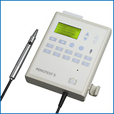
Periotest device
about
Periotest device
References: ... 1989
1990 1991
1992 1993
1994 1995
1996 1997
1998 1999
2000 2001
2002
2003
...........
2003:
Bone density: its influence on implant stability after uncovering.
MORRIS HE, OCHI S, CRUM P, ORENSTEIN I, PLEZIA R.
J Oral Implantol. 2003;29(6):263-9.
Primary implant stability and bone density are variables that have
long beenconsidered to be essential to achieving predictable osseointegration
andlong-term clinical survival. Although the dentist can control most factorsassociated
with implant survival, bone density is the one factor that cannot becontrolled.
Measuring implant stability would assist in determining if animplant has
integrated and is ready for the fabrication of the final prosthesis.Changes
in implant stability in each type of Bone Quality (BQ-1, -2, -3, and-4),
which may occur with time, have not been studied. Such information couldhelp
identify well-integrated implants and identify changes associated withimpending
implant failure. Several studies have used the Periotest instrument
tostudy implant stability. Use of the Periotest implant stability
will be studiedduring each phase of implant treatment for each bone density,
and a range forclinically satisfactory integration will be suggested. Implant
stability changesover time, and the changes are different for each bone
density as the bonesurrounding the nonhydroxyapatite implant becomes denser.
This is clearlydemonstrated in a postmortem histological specimen. The
changes in implantstability (Periotest Values [PTVs]) are more
apparent in BQ-1 and BQ-2 bone andless apparent in BQ-3 and BQ-4 bone.
The Periotest is capable of providingvaluable information concerning
favorable or unfavorable changes in thebone-implant interface after uncovering.
In addition, it can help identify whenan implant is ready to be loaded.
A new range of PTVs (-5 to -2) is suggestedfor monitoring the status of
implants. Implants with PTVs more positive than -2would indicate a bone-implant
complex that may be marginal.
DVA Dental Clinical Research Center, Ann Arbor, MI 48105, USA.
Rotational panoramic versus intraoral rectangular radiographs
for evaluation ofperi-implant bone loss in the anterior atrophic mandible.
ZECHNER W, WATZAK G, GAHLEITNER A, BUSENLECHNER D, TEPPER G, WATZEK
G.
Int J Oral Maxillofac Implants. 2003 Nov-Dec;18(6):873-8.
PURPOSE: In patients with atrophic mandibles, elevation of the floor
of themouth often prevents intraoral rectangular radiography for longitudinalfollow-up
studies, while extraoral techniques such as panoramic radiographs tendto
produce distorted views of the interforaminal region. In this study,intraoral
rectangular radiographs and panoramic radiographs were compared fortheir
accuracy in evaluating peri-implant bone loss. MATERIALS AND METHODS: In
arecall program, 22 patients with 88 screw-type implants (44 MKII and 44
Frios)were followed. Interforaminal marginal bone loss was evaluated by
extraoralorthopantomograms and by intraoral rectangular radiographs. In
addition, pocketdepth, Periotest readings, and bleeding on probing
were recorded. Forstatistical analysis, the Spearman coefficient of correlation
was used. Theeffects on bone loss and clinical variables were computed
with a mixed model andthe Bland and Altman method. RESULTS: Computed as
least square means, the meandifference between panoramic radiographs (2.4
+
0.2 mm for MKII implants and1.6 + 0.2 mm for Frios implants) and
intraoral radiographs (2.6 + 0.2 mm and1.4 + 0.2 mm, respectively)
was 0.2 mm (range, 0.1 to 0.8 mm). DISCUSSION: Inthis study, the 2 imaging
techniques were comparable clinically in terms of theprecision with which
they could be used to measure marginal bone loss.CONCLUSION: For highly
atrophic mandibles with unfavorable imaging conditions,rotational panoramic
radiographs can be a useful alternative to intraoralsmall-format radiographs
for evaluating peri-implant bone loss.
Department of Oral Surgery, Dental School, University of Vienna, LudwigBoltzmann
Institute of Oral Implantology, Vienna, Austria.werner.zechner@univie.ac.at
Four-year follow-up of larger-diameter implants placed
in fresh extractionsockets using a resorbable membrane or a resorbable
alloplastic material.
PROSPER L, GHERLONE EF, REDAELLI S, QUARANTA M.
Int J Oral Maxillofac Implants. 2003 Nov-Dec;18(6):856-64.
Clinical Trial Randomized Controlled Trial
PURPOSE: The aim of this randomized study was to evaluate and compare
thelong-term success rates of cylindric, screw-type titanium implants with
a largerdiameter (5.9 mm) that were placed in fresh extraction sockets
in associationwith resorbable bone substitutes or a resorbable membrane.
MATERIALS ANDMETHODS: Eighty-three partially edentulous adult patients,
selected from amongthose treated in 1997 and 1998 at the San Raffaele Institute
in whom 1 or moreimplants had been placed into fresh posterior mandibular
or maxillary sockets,were included in the study. A total of 111 implants
were placed, 36 in mandiblesand 75 in maxillae. Fifty-six implants were
placed in combination withresorbable hydroxyapatite (HA group) and 55 with
a resorbable membrane (MRgroup). Intraoral radiographs and follow-up examinations,
including verificationof implant stability via the Periotest,
were carried out at second-stage surgery3, 6, 9, and 12 months later; and
then annually up to 4 years after placement ofthe definitive restoration.
The radiographic examination was conducted by meansof a standardized procedure
to verify osseointegration. RESULTS: There was 100%attendance at the follow-up
examination after 4 years. At second-stage surgery,which was performed
after 4 to 6 months' healing time, none of the implantsshowed any signs
of mobility, peri-implantitis, or bone loss. Two implantsfailed in the
MR group, one at 3 months and one at 9 months after placement; 1implant
failed in the HA group at 4 months after placement. After 4 years, theimplant
success rate was 97.3% (108 of 111 implants were considered successful).The
success rate did not differ significantly between the HA group (98.2%)
andthe MR group (96.4%). DISCUSSION: The use of larger-diameter implants
served tominimize the anatomic discrepancies that would have evolved when
substituting amolar with a standard-diameter implant. According to the
accepted criteria forsuccess, the 5-year success rate should be at least
85%; therefore both methodsmay be considered satisfactory. CONCLUSION:
Implants placed in combination witha resorbable allogeneic material or
with a resorbable membrane providedpredictable long-term results when restored
with a fixed partial denture.
Oral Rehabilitation Department, University Vita e Salute, Scientific
Institute,San Raffaele Hospital, Milan, Italy. lorisprosper@tin.it
Randomized multicenter comparison of 2 IMZ and 4 TPS
screw implants supportingbar-retained overdentures in 425 edentulous mandibles.
MAU J, BEHNEKE A, BEHNEKE N, FRITZEMEIER CU, GOMEZ-ROMAN G, D'HOEDT
B,SPIEKERMANN H, STRUNZ V, YONG M.
Int J Oral Maxillofac Implants. 2003 Nov-Dec;18(6):835-47.
Clinical Trial Multicenter Study
Randomized Controlled Trial
PURPOSE: Two treatment concepts for implant-supported bar retention
ofmandibular overdentures-2 intramobile cylinder (IMZ) implants and a Dolder
barand 4 titanium plasma-sprayed (TPS) screw implants and an angulated
bar-werecompared in a randomized controlled clinical trial with respect
topostprosthetic efficacy and safety. MATERIALS AND METHODS: Four hundredtwenty-five
patients with edentulous mandibles were enrolled; 212 wererandomized to
TPS implants (control group) and 213 to IMZ implants (test group).Endpoints
were occurrences of postprosthetic integration deficiency (ID),functional
deficiency (FD), and complications. The trial was sized to detect a10%
difference in 5-year ID-free postprosthetic system lifetime with a power
of80%. RESULTS: With 340 protocol-completed cases, the trial achieved itspredetermined
power. The 2 systems did not show statistically significantdifferences
in occurrences of postprosthetic ID and FD; 5-year occurrence-freepostprosthetic
system lifetime probabilities were estimated as 42.5% with IMZand 42.8%
with TPS, for ID; and as 82.6% with IMZ and 87.2% with TPS, for FD.However,
at 3 to 6 months after surgery, mean Periotest values weresignificantly
higher (P = .0001 without adjustment) with IMZ implants (5.6, SD4.2) than
with TPS implants (0.8, SD 4.3). TPS implants showed a higherincidence
of inflammation and recession, while IMZ implants had a higherincidence
of implant fracture after functional loading. DISCUSSION: Thesystem-wise
approach overcomes potential bias with implant-wise analyses. Acombination
of radiographic and clinical criteria distinguishes betweendesirable integration
and functional anchorage. The in situ survival rates at 5years in this
study (95% for IMZ, 92% for TPS) match rates reported in theliterature.
CONCLUSION: This study demonstrated equivalent efficacy of 2 IMZcylinders
and 4 TPS screws in implant-supported, bar-retained mandibularoverdentures
and indicated a higher rate of complications with the TPS screwimplants.
Department of Statistics in Medicine, Heinrich Heine University, Duesseldorf,Germany.
ismmau@uni-duesseldorf.de
Dental implants in reconstructed jaws: implant longevity
and peri-implant tissueoutcomes.
CHEUNG LK, LEUNG AC.
J Oral Maxillofac Surg. 2003 Nov;61(11):1263-74.
PURPOSE: The study aimed to evaluate the clinical status and survival
of dentalimplants inserted in reconstructed jaws, with particular reference
to theperi-implant tissues. MATERIALS AND METHODS: We conducted a clinical
follow-upstudy based on 29 rehabilitated patients after oral tumor surgery,
who receivedautogenous bone grafts from the ilium and endosseous implants
(14 maxillary and15 mandibular cases; 140 implants) for functional jaw
reconstruction between1988 and 1999. Clinical records of the patients were
reviewed retrospectively.Clinical parameters of plaque index, probing pocket
depth, and bleeding onprobing were assessed around the implants and control
teeth at 4 locations(mesiobuccal, distobuccal, mesiolingual, and distolingual).
Implant mobility wasassessed clinically and objectively using a Periotest
(Gulden; Siemens,Bensheim, Germany [www.med-gulden.com]) equipment for
those implants supporting removable prostheses.Radiographically, the proportion
of implant length remained osseointegrated wasmeasured. RESULTS: With a
mean follow-up time of 50 months, 90.7% of the 140implants placed were
functional in supporting dental prostheses; 4.3% ofimplants failed in osseointegration
and the remaining 5.0% implants wereosseointegrated but nonfunctional.
A total of 493 sites of 127 functionalimplants and 392 sites of 98 control
teeth were assessed. No significantdifference was found between the implants
and control teeth parameters, excepton the probing pocket depth. The mean
peri-implant probing depth was 3.5 mm, and52.7% of the measured sites were
3 mm or less. More than one third of theimplants (35.9%) presented with
increased probing depth (> or =4 mm), and thiswas significantly higher
than in the control teeth (P <.001, chi(2) test).Bleeding on probing
was found in 19.3% of the measured peri-implant sites,corresponding to
42.2% of the dental implants. Of the implants, 28.9% werecompletely free
from plaque and 9.4% show visible plaque accumulation. Mobilityassessment
was feasible on 32 implants and no mobility was detected.Radiographically,
the mean implant length remained in bone was 81.1%, with 82.6%in the maxilla
and 79.4% in the mandible. Implant survival rate calculated usingthe Kaplan-Meier
method was 86.9% for 5 years. Based on the defined criteria,the success
rate of implants placed in reconstructed jaws in this study was90.7%. CONCLUSION:
Endosseous implants can be successfully placed inreconstructed jaws for
oral rehabilitation with maintenance of reasonable healthstatus of the
peri-implant tissues in the long-term.
Oral and Maxillofacial Surgery, Prince Philip Dental Hospital, 34 Hospital
Road,Hong Kong SAR, China. lkcheung@hkucc.hku.hk
Implant stability and histomorphometry: a correlation
study in human cadavers using stepped cylinder implants
NKENKE E, HAHN M, WEINZIERL K, RADESPIEL-TROGER M, NEUKAM FW, ENGELKE
K
Clin Oral Implants Res 14(5):601-609 (2003)
The aim of the present study was to determine the correlation between
the primary stability of dental implants placed in edentulous maxillae
and mandibles, the bone mineral density and different histomorphometric
parameters. After assessing the bone mineral density of the implant sites
by computed tomography, 48 stepped cylinder screw implants were installed
in four unfixed human maxillae and mandibles of recently deceased people
who had bequeathed their bodies to the Anatomic Institute I of the University
of Erlangen-Nuremberg for medical-scientific research. Peak insertion torque,
Periotest
values and resonance frequency analysis were assessed. Subsequently, histologic
specimens were prepared, and bone-to-implant contact, the trabecular bone
pattern factor (TBPf), the density of trabecular bone (BV/TV) and the height
of the cortical passage of the implants were determined. The correlation
between the different parameters was calculated statistically. The mean
resonance frequency analysis values (maxilla 6130.4+363.2 Hz, mandible
6424.5+236.2 Hz) did not correlate with the Periotest measurements
(maxilla 13.1+7.2, mandible -7.9+2.1) and peak insertion
torque values (maxilla 23.8+2.2 N cm, mandible 45.0+7.9 N
cm) (P=0.280 and 0.193, respectively). Again, no correlations could be
found between the resonance frequency analysis, the bone mineral density
(maxilla 259.2+124.8 mg/cm3, mandible 349.8+113.3 mg/cm3),
BV/TV (maxilla 19.7+8.8%, mandible 34.3+6.0%) and the TBPf
(maxilla 2.39+1.46 mm-1, mandible -0.84+3.27 mm-1) (P=0.140
and 0.602, respectively). However, the resonance frequency analysis values
did correlate with bone-to-implant contact of the oral aspect of the specimens
(maxilla 12.6+/6.0%, mandible 35.1+5.1%) and with the height
of the crestal cortical bone penetrated by the implants in the oral aspect
of the implant sites (maxilla 2.1+0.7 mm, mandible 5.1+3.7
mm) (P=0.024 and 0.002, respectively). The Periotest values showed
a correlation with the height of the crestal cortical bone penetrated by
the implants in the buccal aspect of the implant sites (maxilla 2.5+1.2
mm, mandible 5.4+1.2 mm) (P=0.015). The resonance frequency analysis
revealed more correlations to the histomorphometric parameters than the
Periotest
measurements. However, it seems that the noninvasive determination of implant
stability has to be improved in order to give a more comprehensive prediction
of the bone characteristics of the implant site.
Provisional implants for anchoring removable interim prostheses
in edentulous jaws: a clinical study.
KRENNMAIR G, WEINLANDER M, SCHMIDINGER S (Vienna, Austria)
Int J Oral Maxillofac Implants 18(4):582-8 (2003)
Dental Implants, Dental Prosthesis, Dental Restoration, Follow-Up
Studies, Human, Middle Age, Patient Satisfaction
PURPOSE: The behavior of provisional implants in edentulous maxillae/mandibles
used for anchoring removable interim overdentures was followed for the
time of the intended healing of the definitive implants. MATERIALS AND
METHODS: Twenty-eight edentulous arches (19 maxillae, 9 mandibles) were
provided with 77 provisional implants (2 to 4 in maxillae; 2 or 3 in mandibles)
for anchoring removable interim prostheses (overdentures). The provisional
implants were to be maintained until final restoration (6 to 9 months in
the maxilla and 3 months in the mandible). The loss rate of provisional
implants and handling and behavior of the anchored overdenture were monitored
until the definitive prosthetic restoration was placed. RESULTS: Twenty-three
(29.8%) of the 77 provisional implants were lost prematurely. The loss
rate of maxillary provisional implants (21/58; 36.2%) was significantly
higher than that of mandibular implants (2/19; 10.5%) (P < .01). Determination
of terminal stability (by means of the Periotest) of the provisional
implants showed higher stability in the mandible (+3.8 +2.3) than
in the maxilla (+8.6 + 3.9) (P < .05). In obvious contrast to
mandibular interim overdentures, handling of maxillary interim overdentures
was found to improve significantly during the follow-up period (P <
.01). DISCUSSION AND CONCLUSION: With both the low loss rate in the mandible
and the higher loss rate seen in the maxilla, placement of provisional
implants fulfills the requirements for initiating immediate prosthetic
rehabilitation. The removable interim overdenture can be adequately stabilized
and provides for added patient comfort and satisfaction as compared to
a conventional complete denture. An important aspect of the continued use
of provisional implants concerns the expectations placed in these implants
by both clinician and patient, which are quite different than those for
definitive implants.
Restoration of partially edentulous patients using dental implants
with a microtextured surface: a prospective comparison of delayed and immediate
full occlusal loading
CANNIZZARO G, LEONE M (Italy)
Int J Oral Maxillofac Implants 18(4):512-22 (2003)
Comparative Study, Crowns, Dental Implants, Dental Prosthesis, Follow-Up
Studies, Human, Middle Age, Radiography, Time Factors, Treatment Outcome
PURPOSE: The aim of this study was to determine the clinical effectiveness
of placing dental implants with microtextured surfaces into full occlusal
loading at the time of placement in partially edentulous patients. MATERIALS
AND METHODS: Two demographically similar groups of 14 patients each were
treated with a total of 92 Spline Twist Implants (Centerpulse Dental, Carlsbad,
CA). Test implants were placed into immediate full occlusal loading, and
control implants were restored using a conventional delayed loading procedure.
Otherwise, both groups of patients received similar therapy from the same
treatment team. Radiographs, periodontal indices, and Periotest
values were recorded every 6 months during routine clinical follow-up appointments.
The mean loading time for all prostheses was 24 months at the time of this
report. RESULTS: No implants failed in the test group, and 1 implant failed
before loading in the control group. Cumulative implant success was 98.9%
for all implants placed (test group = 100%; control group = 92.9%). Periodontal
measurements indicated no significant clinical differences between implants
placed into immediate full occlusal loading and those loaded via a conventional
delayed protocol. DISCUSSION: Immediate full occlusal loading of partial
prostheses supported by microtextured implants in partially edentulous
patients demonstrated excellent clinical results, with no adverse periodontal
effects after 24 months of function. Additional follow-up will provide
invaluable information on the long-term effects of this technique. CONCLUSION:
Immediate full occlusal loading of partial prostheses supported by microtextured
implants can be successfully achieved for 24 months in highly motivated
patients with excellent oral hygiene.
The Periotest in traumatology. Part II. The Periotest as a special
test for assessing the periodontal status of teeth in children that have
suffered trauma
ANDRESEN M, MACKIE I, WORTHINGTON H (Manchester, UK.)
Child, Comparative Study, Follow-Up Studies, Human, Incisor, Percussion,
Periapical Diseases, Root Resorption, Sound/diagnostic use, Tooth Fractures,
Tooth Mobility, radiography, Wound Healing
Dent Traumatol 19(4):218-20 (2003)
The research was carried out on the upper permanent incisor teeth of
101 children aged 8-12 years attending the trauma clinic at the University
Dental Hospital, Manchester, UK. The double blind, cross-over study involved
Periotest
readings being taken by one examiner and traditional special tests being
carried out by another examiner. The traditional special tests chosen were
mobility testing, percussion sounds and radiographs. The Periotest
readings and special tests were then repeated at the child's recall appointment.
The results revealed that there was a significant difference (P < 0.001)
between the number of abnormalities detected by the Periotest
and those detected by the traditional special tests. There was also found
to be a significant difference (P < 0.001) in the detection of undesirable
sequelae between the Periotest and the traditional special tests.
The Periotest in traumatology. Part I. Does it have the properties
necessary for use as a clinical device and can the measurements be interpreted?
ANDRESEN M, MACKIE I, WORTHINGTON H (Manchester, UK.)
Dent Traumatol 19(4):214-7 (2003)
Child, Cooperative Behavior, Human, Incisor, Observer Variation,
Periodontics, Reproducibility of Results, Time Factors
The Periotest is a non-invasive, electronic device which provides
an objective measurement of the reaction of the periodontium to a defined
impact load applied to the tooth crown. In order for a clinician to interpret
Periotest
readings, normal physiological values must be known. In traumatology, using
the contralateral tooth as a control is impractical, as it too may be affected
by injury. The main purpose of this research project was to establish the
normal physiological Periotest readings for upper permanent incisor
teeth at an age when trauma to teeth is at a peak. This was achieved by
taking Periotest readings on healthy upper permanent incisors
of 250 children aged between 8-12 years. Test-retest reliability was investigated
by taking repeated Periotest readings on the teeth of 13 dental
nurses at timed intervals over a 2-h period. The practicality of using
the device on children was examined by examiner observation and patient
questionnaire. Results showed a wide variation of Periotest readings
in healthy upper permanent incisor teeth in 8-12-year-olds, and thus it
was felt that normal physiological measurements are best expressed as a
range. The test-retest reliability of readings was good (R > 0.85) provided
that the repeat reading was taken at least 15 min after the initial reading.
The examiner found the equipment easy to use and taking measurement was
readily acceptable by the sample group. It was concluded that Periotest
could be utilised in traumatology, although the clinician needs to take
into account the limitations of the device and the difficulty in interpreting
the results.
In-patient comparison of immediately loaded and non-loaded implants
within 6 months.
LORENZONI M, PERTL C, ZHANG K, WEGSCHEIDER WA (Graz, Austria)
Clin Oral Implants Res 14(3):273-9(2003)
Alveolar Process, Bone Resorption, Comparative Study, Dental Abutments,
Dental Implants, Follow-Up Studies, Human, Middle Age, Osseointegration,
Prospective Studies, Time Factors, Treatment Outcome, Wound Healing
According to the Branemark protocol, a stress-free healing period is
one of the most emphasised requirements for implant integration. Recent
studies have encouraged a progressive shortening of the healing period
and immediate loading has been proposed for the edentulous mandible. This
prospective study evaluated the clinical outcomes of 14 immediately loaded
FRIALIT-2(R) implants compared with 28 non-loaded controls in an in-patient
study. The results were based on clinical stability and on changes of bone
level from implant placement to abutment connection 6 months after insertion.
In the course of our investigation, seven patients with edentulous mandibles
have been treated with 43 implants following an immediate-loading protocol.
Six FRIALIT-2(R) implants were placed in the interforaminal region located
at positions 34, 33, 32, 42, 43, 44. Bone level in relation to implant
margin was measured and recorded. In order to obtain an in-patient comparison
of immediately loaded and non-loaded implants, the ones at 33 and 43 were
chosen to be immediately loaded by a Dolder-bar retained overdenture. The
implants in position 32, 34, 42 and 44 were covered and left to heal. After
a healing period of 6 months, second stage surgery was carried out. The
clinical criteria to be checked at this point were survival, Periotest
values and marginal bone level at the loaded and non-loaded implants. The
mean Periotest value was -2.7 for the loaded and -5.6 for the
non-loaded implants. The Mann-Whitney U-test showed that the difference
was highly significant (P < 0.001). The mean bone level changes at prosthetic
delivery were 0.9 mm resorption for the loaded implants and 0.33 mm for
non-loaded implants. The difference was highly significant (P < 0.001).
No implant failures were observed up to the prosthetic restoration 6 months
post insertion. The results of this investigation allowed for direct comparison
of implant survival and clinical results between immediately loaded implants
and standard implants. Clinical bone changes at the 6-month evaluation
demonstrated significantly higher crestal resorption around loaded implants.
This fact was confirmed by higher median Periotest values (-3
vs. -6) of immediately loaded implants. According to the outcome of this
study, immediate loading of two interforaminal implants with a Dolder-bar
resulted in an intimate bone apposition comparable with implants with submerged
healing. Nevertheless, the coronal bone level as well as clinical stability
(PTV) were significantly lower in the case of the immediately loaded implants.
Future studies will be necessary to evaluate marginal bone resorption,
Periotest
values and clinical success rates of mandibular immediately loaded implants
in the long-term.
A split-mouth study on periodontal and microbial parameters in children
with complete unilateral cleft lip and palate.
QUIRYNEN M, DEWINTER G, HEIDBUCHEL K, VERDONCK A, CARELS C (Leuven,
Belgium)
J Clin Periodontol 30(1):49-56(2003)
Alveolar Bone Loss, Child, Cleft Lip therapy, Comparative Study,
Dental Plaque Index, Follow-Up Studies, Gingival Hemorrhage, Gingival Recession,
Gram-Negative Bacteria, Human, Periodontal Diseases, Tooth Mobility
BACKGROUND: Complete unilateral cleft lip and palate (UCLP) is a hereditary
or multifactorial malformation that can be corrected successfully with
a combined orthodontic, surgical and restorative treatment. Such multidisciplinary
treatment takes many years and demands a lot of attention to both patients'
teeth and periodontium. OBJECTIVES: This split-mouth study aimed to compare
the periodontal health as well as the microbial parameters between cleft
and non-cleft region. MATERIAL AND METHODS: 75 patients (52 males, 23 females)
between 8 and 20 years with a complete unilateral cleft lip and palate
(before (n = 30), during (n = 34) and after (n = 11) the active orthodontic
treatment) volunteered for this study. Four regions were defined for the
split-mouth comparison: teeth neighbouring cleft (site 1), tooth in cleft
(site 2), and the corresponding contra-lateral teeth, respectively, in
the unaffected quadrants (sites 3 and 4). At all sites the following periodontal
parameters were recorded: plaque and gingivitis indices, pocket depth,
attachment loss, bleeding on probing, tooth mobility (visual and Periotest),
radiographic bone loss and gingival width. In addition, three pooled subgingival
plaque samples were taken (around tooth in cleft, teeth facing the cleft,
and contra-lateral teeth of the latter). RESULTS: The differences between
the teeth neighbouring the cleft and the corresponding contra-lateral opponents
were of borderline significance (P <or= 0.05) for the plaque index,
the approximal probing depths and the attachment loss (teeth facing the
cleft always had slightly higher parameters). When the tooth in the cleft
was compared to the contra-lateral tooth, differences were only found for
both the approximal probing depths, attachment loss and bone loss, which
were significantly higher for the tooth in the cleft. The microbial analysis
did not reveal differences between the different sites, neither in the
proportion of aerobic and anaerobic bacteria (differences < 0.5 log),
nor in the detection frequency of periopathogens. CONCLUSIONS: These data
indicate that the periodontium in UCLP patients can cope well with a long-term
orthodontic treatment, even in unfavourable conditions (like absence of
attached gingiva and poor oral hygiene).
Evaluation of distraction implants for prosthetic treatment after
vertical alveolar ridge distraction: a clinical investigation.
FEICHTINGER M, GAGGL A, SCHULTES G, KARCHER H (Graz, Austria)
Int J Prosthodont 16(1):19-24 (2003)
Alveolar Bone Loss, Alveolar Ridge Augmentation, Dental Implants,
Dental Prosthesis, Follow-Up Studies, Human, Middle Age, Osseointegration,
Periodontal Pocket
PURPOSE: The purpose of this study was to clinically evaluate the mucosal
condition and the esthetic and functional results of distraction implants
loaded with fixed or removable implant-supported restorations. MATERIALS
AND METHODS: A total of 35 patients were treated with 62 distraction implants
for correction of alveolar ridge deficiency. The distraction implants were
loaded with prosthetic superstructures 4 to 6 months after distraction.
Nine patients were provided with single-crown restorations, 16 received
metal-ceramic fixed partial dentures, and 10 received removable overdentures.
Recall was scheduled before and 3, 6, and 9 months after implant loading.
Periotest
values, periimplant probing depths, and radiographic marginal bone levels
were recorded, along with any biologic or mechanical complications. RESULTS:
Patients were followed for 9 months after implant loading. Two distraction
implants were lost before abutment connection. After fabrication and placement
of individual abutments, all implants were loaded with prosthetic superstructures.
The results showed a decrease of the Periotest values, and thus
an increase of implant stability, during the following 9 months. Periimplant
probing depths also decreased in the first months after implant loading.
Soft tissue around the superstructures and adjacent teeth was healthy.
CONCLUSION: The distraction implant system has a high potential for osseointegration.
Because of the gentle distraction technique and the possibility of using
individual abutments at almost any angulation, satisfying esthetic and
functional results are possible. The rate of complications was low in this
short-term study.
Immediate loading of single-tooth implants in the anterior maxilla.
Preliminary results after one year.
LORENZONI M, PERTL C, WIMMER G, WEGSCHEIDER WA (Graz, Austria)
Clin Oral Implants Res 14(2):180-7 (2003)
Acrylic Resins, Adult, Alveolar Process, Bone Resorption, Crowns,
Dental Abutments, Dental Implants, Single-Tooth, Dental Restoration, Temporary,
Follow-Up Studies, Human, Incisor, Middle Age, Occlusal Splints, Osseointegration,
Survival Analysis, Treatment Outcome, Wound Healing
According to the standard protocol, a load-free healing period is one
of the most emphasized requirements for implant integration. Recent studies
have encouraged a progressive shortening of the healing period for single-tooth
implants and immediate loading has been proposed for the aesthetic zone
in the maxilla. The present study evaluated clinical outcomes of immediately
loaded FRIALIT-2 Synchro implants 12 months after placement in the maxillary
incisal region. In the course of our investigation, nine patients have
been treated following an immediate loading protocol. The stepped-screw
type implants were inserted with an increasing torque up to 45 Ncm, thus
measuring the primary stability of the implants. All implants were immediately
restored with unsplinted acrylic resin provisional crowns and the patients
provided with occlusal splints. Regular controls were performed at monthly
intervals, intraoral radiographs were taken directly after implant placement,
6 and 12 months post insertion. The survival rate, clinical stability (Periotest)
and radiographic coronal bone defects (CBD) were evaluated at delivery
of the definitive superstructures (CBD 6) and 6 months later (CBD 12).
Twelve FRIALIT-2 Synchro stepped screws of 3.8, 4.5 and 5.5 mm diameter
and 13 and 15 mm length were placed in the incisal maxillary region. The
median Periotest value 6 months post insertion was -2 with a minimum
of -5 and a maximum of +2. The mean coronal bone level changes (CBD) at
6 and 12 months were 0.45 and 0.75 mm. No implant failed up to 12 months
after insertion, resulting in a 100% survival rate. The presented results
showed promising data for immediately loaded single-tooth implants in the
anterior maxilla. Periotest values were within the range published
for submerged implants. The radiographic coronal bone resorption after
6 and 12 months was even less than evaluated for implants placed in a standard
two-stage procedure. It is evident that successful immediate loading protocols
require a careful and strict patient selection aimed at achieving the best
primary stability and avoiding any excessive functional or non-functional
loading. Additional research needs to be done to provide data in situations
where problems of poor bone quality, multiple implants or augmentation
procedures must be overcome.
References: ... 1989
1990 1991
1992 1993
1994 1995
1996 1997
1998 1999
2000 2001
2002
2003
...........
YOUR COMMENTS, CRITICAL REMARKS OR SUGGESTIONS ARE VERY HELPFUL!
-
 Dieter Lukas,
update: 2004-VIII-21.